Copyright
©The Author(s) 2022.
World J Clin Cases. Feb 16, 2022; 10(5): 1689-1696
Published online Feb 16, 2022. doi: 10.12998/wjcc.v10.i5.1689
Published online Feb 16, 2022. doi: 10.12998/wjcc.v10.i5.1689
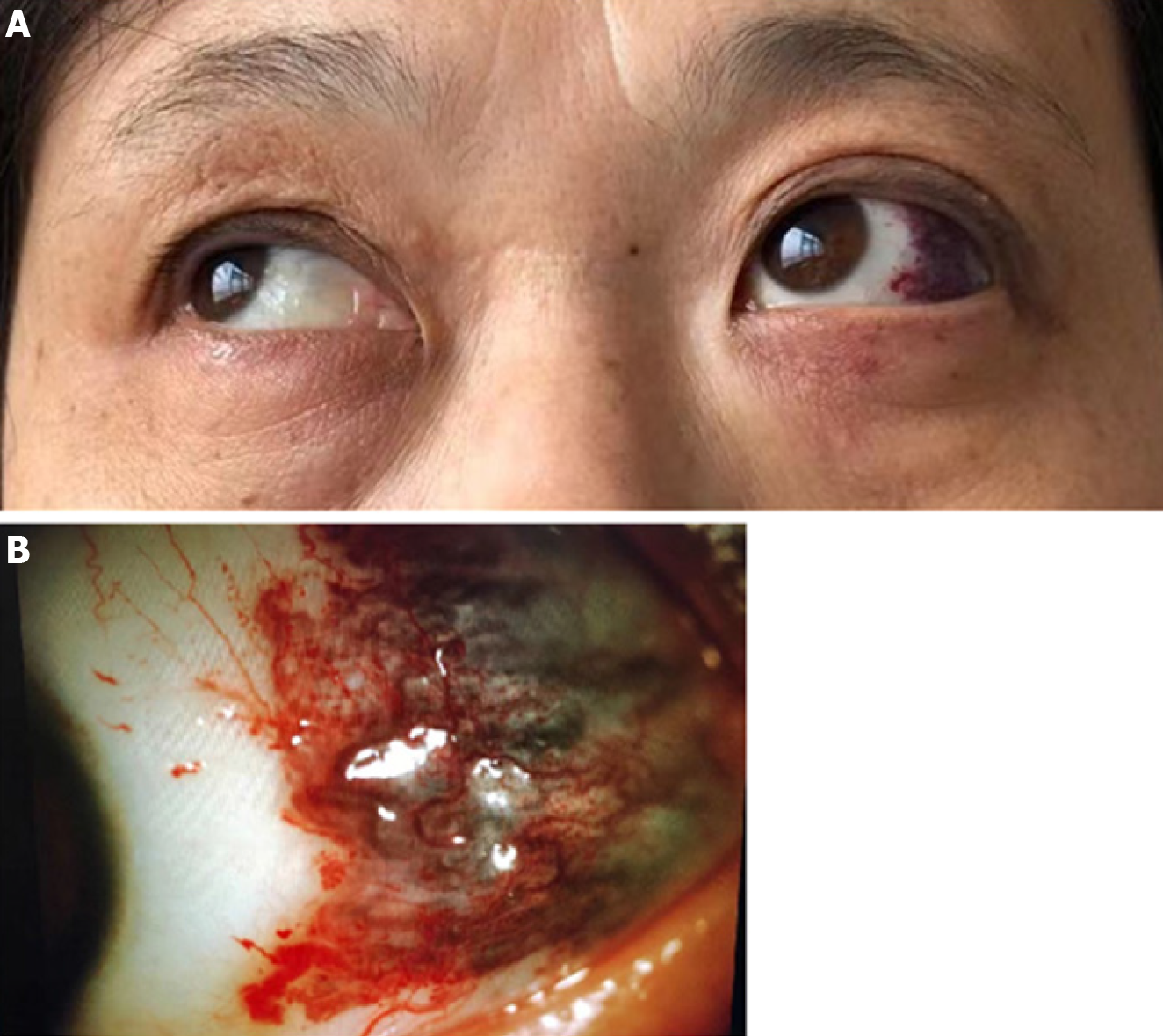
Figure 1 The slit-lamp examination of left eye.
A: The left eyeball protruded forward; B: The slit-lamp examination found that her left conjunctiva was not congestible and curled blood vessels were seen under the conjunctiva in the temporal side, which color was dark purple, with a range of about 1 cm × 1 cm.
- Citation: Lei JY, Wang H. Bulbar conjunctival vascular lesion combined with spontaneous retrobulbar hematoma: A case report. World J Clin Cases 2022; 10(5): 1689-1696
- URL: https://www.wjgnet.com/2307-8960/full/v10/i5/1689.htm
- DOI: https://dx.doi.org/10.12998/wjcc.v10.i5.1689


