Copyright
©The Author(s) 2022.
World J Clin Cases. Feb 16, 2022; 10(5): 1645-1653
Published online Feb 16, 2022. doi: 10.12998/wjcc.v10.i5.1645
Published online Feb 16, 2022. doi: 10.12998/wjcc.v10.i5.1645
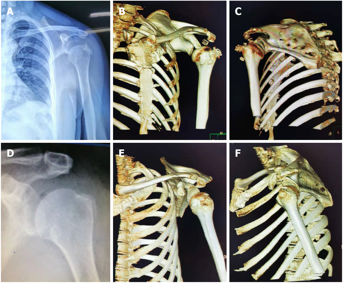
Figure 2 Imaging examinations of case two.
A–C: Plain radiographs and three-dimensional computed tomography (3D-CT) reconstruction of the left shoulder, showing multiple intra-articular loose bodies and the subluxation of the humeral head; D–F: Postoperative radiographic re-examination and 3D-CT reconstruction showed no loose bodies in the subacromial space. The humeral head returned to a normal anatomical relationship.
- Citation: Tang XF, Qin YG, Shen XY, Chen B, Li YZ. Arthroscopic surgery for synovial chondroma of the subacromial bursa with non-traumatic shoulder subluxation complications: Two case reports. World J Clin Cases 2022; 10(5): 1645-1653
- URL: https://www.wjgnet.com/2307-8960/full/v10/i5/1645.htm
- DOI: https://dx.doi.org/10.12998/wjcc.v10.i5.1645


