Copyright
©The Author(s) 2022.
World J Clin Cases. Feb 16, 2022; 10(5): 1572-1579
Published online Feb 16, 2022. doi: 10.12998/wjcc.v10.i5.1572
Published online Feb 16, 2022. doi: 10.12998/wjcc.v10.i5.1572
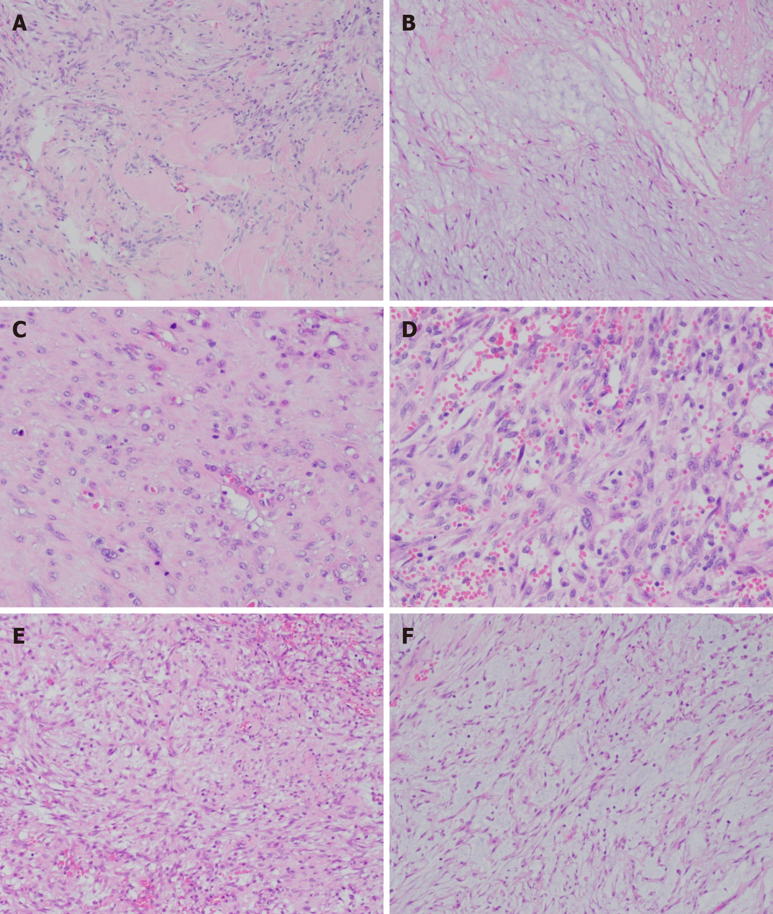
Figure 2 Hematoxylin-eosin staining.
A: Localized fibrous tissue hyperplasia and hyaline degeneration [hematoxylin and eosin (HE), × 100]; B: Some areas showed extracellular mucoid matrix (HE, × 100); C: Mitotic figures (HE, × 200); D: Tumor cells are abundant and there is apparent extravasation of red blood cells (HE, × 200); E: Spindle-shaped and fibroblast-like tumor cells (HE, × 100); F: Spindle-shaped tumor cells with stromal mucous degeneration (HE, × 100).
- Citation: Yu SL, Sun PL, Li J, Jia M, Gao HW. Giant nodular fasciitis originating from the humeral periosteum: A case report. World J Clin Cases 2022; 10(5): 1572-1579
- URL: https://www.wjgnet.com/2307-8960/full/v10/i5/1572.htm
- DOI: https://dx.doi.org/10.12998/wjcc.v10.i5.1572


