Copyright
©The Author(s) 2022.
World J Clin Cases. Feb 6, 2022; 10(4): 1441-1446
Published online Feb 6, 2022. doi: 10.12998/wjcc.v10.i4.1441
Published online Feb 6, 2022. doi: 10.12998/wjcc.v10.i4.1441
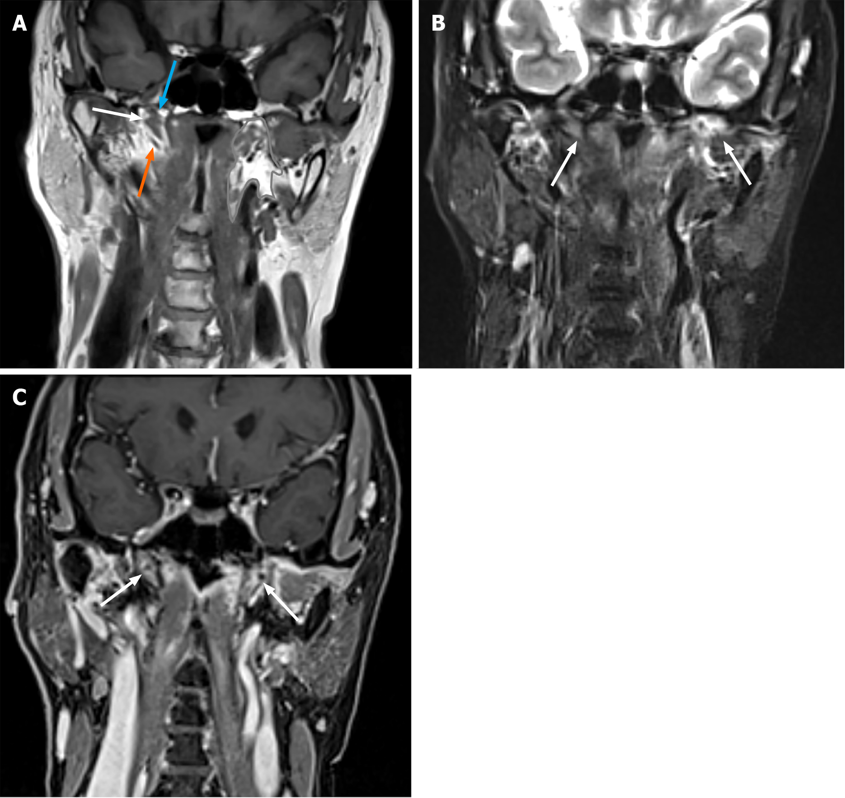
Figure 2 Non-contrast-enhanced and contrast-enhanced images of the patient.
A: Non-contrast-enhanced T1-weighted image of the patient. The Eustachian tube (white arrow) is located in the parapharyngeal space (contoured area). The Merkmal of the Eustachian tube is the levator veli palatine muscle (blue arrow) on the upper side and tensor veli palatine muscle (orange arrow) on the lower side; B: Non-contrast-enhanced fat-saturated T2-weighted image of the patient demonstrated edematous bilateral eustachian tube (white arrow); C: Contrast-enhanced 3D-volumetric interpolated breath-hold examination T1-weighted image demonstrated enhanced bilateral Eustachian tube (white arrow).
- Citation: Yunaiyama D, Aoki A, Kobayashi H, Someya M, Okubo M, Saito K. Eustachian tube involvement in a patient with relapsing polychondritis detected by magnetic resonance imaging: A case report. World J Clin Cases 2022; 10(4): 1441-1446
- URL: https://www.wjgnet.com/2307-8960/full/v10/i4/1441.htm
- DOI: https://dx.doi.org/10.12998/wjcc.v10.i4.1441


