Copyright
©The Author(s) 2022.
World J Clin Cases. Feb 6, 2022; 10(4): 1333-1340
Published online Feb 6, 2022. doi: 10.12998/wjcc.v10.i4.1333
Published online Feb 6, 2022. doi: 10.12998/wjcc.v10.i4.1333
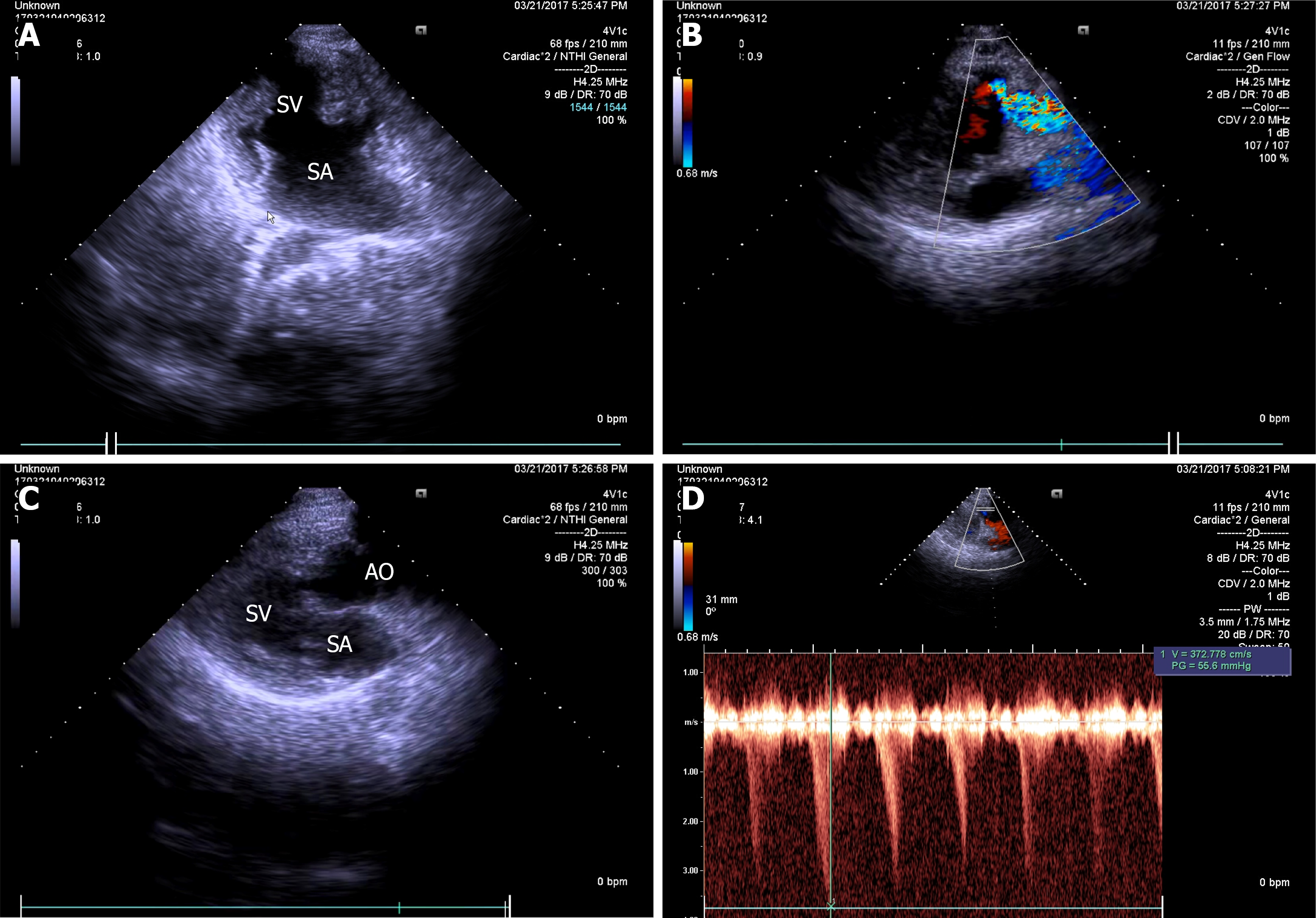
Figure 1 Two-dimensional echocardiographic apical 4-chamber view.
A: Common atrium, single ventricle; B: Hypoplastic pulmonary artery arising from single ventricle; C: Aorta arising from single ventricle; D: Continuous wave Doppler at pulmonary valve with gradient of 55.6 mmHg. SV: Single ventricle; SA: Single atrium; AO: Aorta; PA: Pulmonary artery.
- Citation: Duan HZ, Liu JJ, Zhang XJ, Zhang J, Yu AY. Multiple miscarriages in a female patient with two-chambered heart and situs inversus totalis: A case report . World J Clin Cases 2022; 10(4): 1333-1340
- URL: https://www.wjgnet.com/2307-8960/full/v10/i4/1333.htm
- DOI: https://dx.doi.org/10.12998/wjcc.v10.i4.1333


