Copyright
©The Author(s) 2022.
World J Clin Cases. Feb 6, 2022; 10(4): 1278-1285
Published online Feb 6, 2022. doi: 10.12998/wjcc.v10.i4.1278
Published online Feb 6, 2022. doi: 10.12998/wjcc.v10.i4.1278
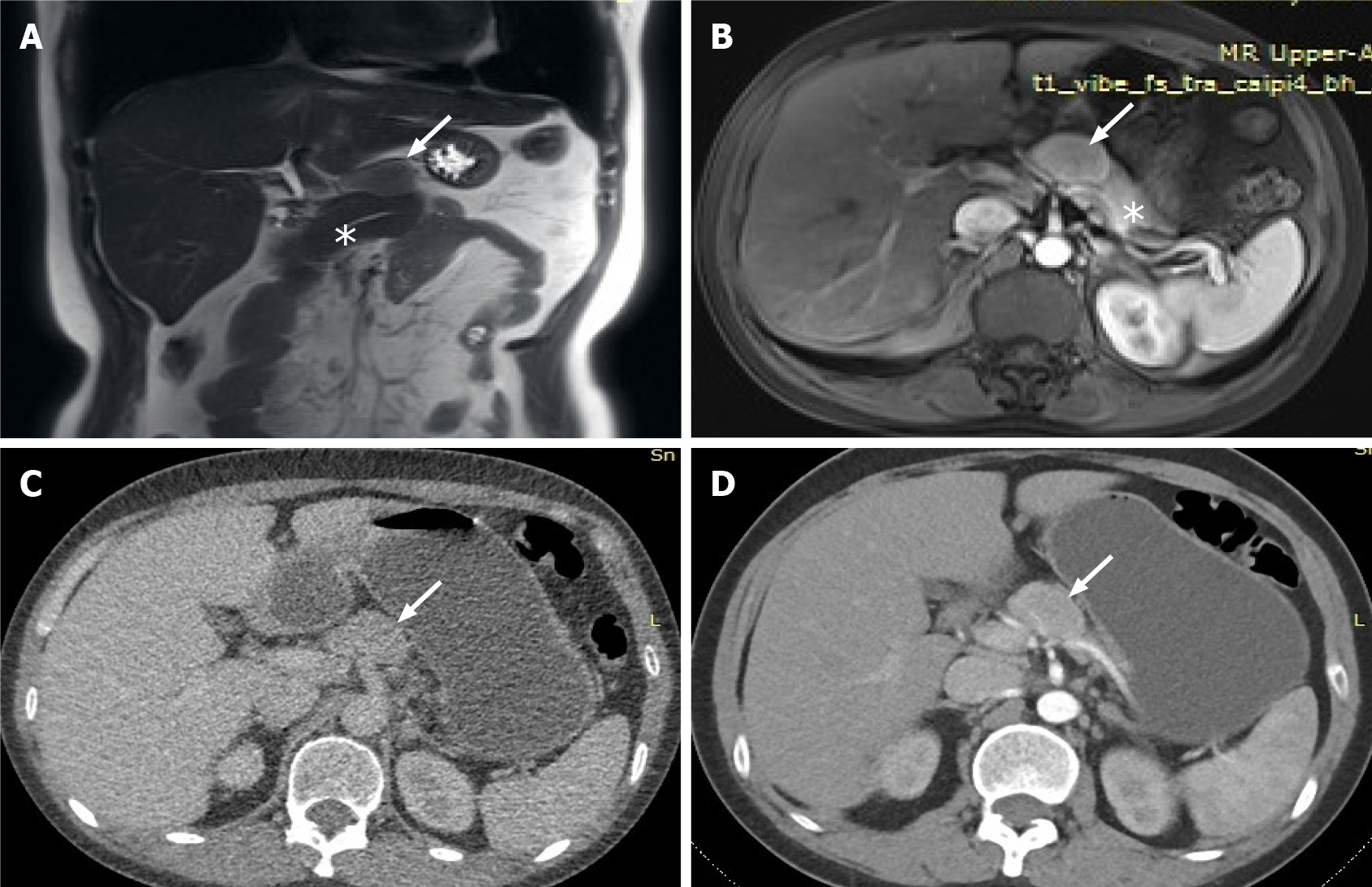
Figure 2 The lesion was further evaluated by computed tomography and magnetic resonance imaging.
A: Magnetic resonance imaging (MRI) showed a lesion of isointensity (white arrow) located above the pancreas body (stellate) which was clearly separated from the normal pancreas; B: Contrast-enhanced -MRI revealed that the edge was enhanced in the arterial phase, and the degree of internal enhancement in each phase was lower than that of the pancreatic parenchyma; C: Computed tomography (CT) showed a nodule of mixed density (white arrow) located above the pancreatic body, which was convex and shallowly lobulated, measuring approximately 37 mm 25 mm in maximum dimensions; D: Contrast-enhanced CT showed uneven progressive enhancement, and the degree of enhancement in each stage was lower than that of pancreatic parenchyma.
- Citation: Zhai HY, Zhu XY, Zhou GM, Zhu L, Guo DD, Zhang H. Unicentric Castleman disease was misdiagnosed as pancreatic mass: A case report. World J Clin Cases 2022; 10(4): 1278-1285
- URL: https://www.wjgnet.com/2307-8960/full/v10/i4/1278.htm
- DOI: https://dx.doi.org/10.12998/wjcc.v10.i4.1278


