Copyright
©The Author(s) 2022.
World J Clin Cases. Feb 6, 2022; 10(4): 1278-1285
Published online Feb 6, 2022. doi: 10.12998/wjcc.v10.i4.1278
Published online Feb 6, 2022. doi: 10.12998/wjcc.v10.i4.1278
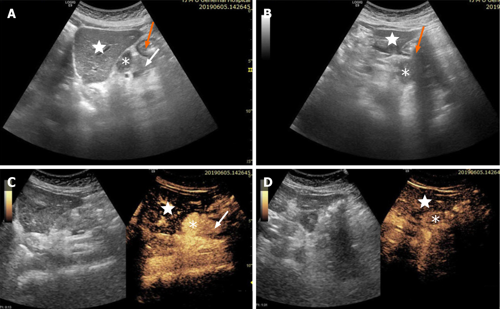
Figure 1 Patient underwent contrast-enhanced ultrasound examination.
Conventional and contrast-enhanced ultrasound of the hypoechoic mass (stellate) between the body of the pancreas (white arrow), left lobe of the liver (white star) and stomach (orange arrow), which has clear boundaries, irregular shape, uneven echoes, and no obvious blood flow signals. The hypoechoic mass showed homogeneous hyperenhancement in the arterial phase, and slightly high enhancement in the venous phase, and measured approximately 56 mm × 37 mm × 25 mm.
- Citation: Zhai HY, Zhu XY, Zhou GM, Zhu L, Guo DD, Zhang H. Unicentric Castleman disease was misdiagnosed as pancreatic mass: A case report. World J Clin Cases 2022; 10(4): 1278-1285
- URL: https://www.wjgnet.com/2307-8960/full/v10/i4/1278.htm
- DOI: https://dx.doi.org/10.12998/wjcc.v10.i4.1278


