Copyright
©The Author(s) 2022.
World J Clin Cases. Dec 26, 2022; 10(36): 13451-13457
Published online Dec 26, 2022. doi: 10.12998/wjcc.v10.i36.13451
Published online Dec 26, 2022. doi: 10.12998/wjcc.v10.i36.13451
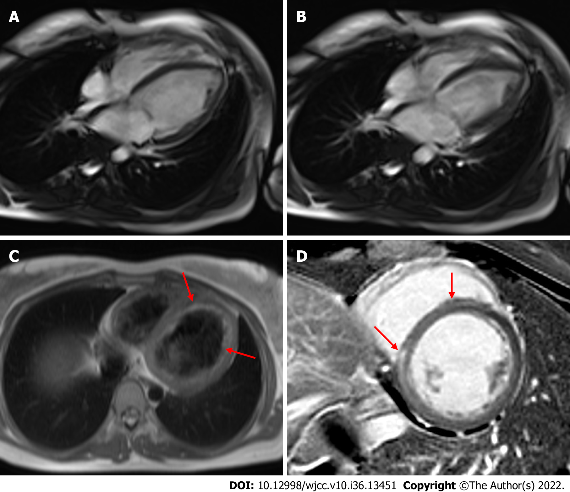
Figure 3 Cardiac magnetic resonance.
A and B: Decreased left ventricular systolic function was observed (A: Diastole and B: Systole); C: A T2-weighted image shows diffuse endocardial edema (arrow); D: A T1-delayed image revealed late gadolinium enhancement in the mid-layer of the interventricular septum (arrow).
- Citation: Lee SD, Lee HJ, Kim HR, Kang MG, Kim K, Park JR. Development of dilated cardiomyopathy with a long latent period followed by viral fulminant myocarditis: A case report. World J Clin Cases 2022; 10(36): 13451-13457
- URL: https://www.wjgnet.com/2307-8960/full/v10/i36/13451.htm
- DOI: https://dx.doi.org/10.12998/wjcc.v10.i36.13451


