Copyright
©The Author(s) 2022.
World J Clin Cases. Dec 26, 2022; 10(36): 13435-13442
Published online Dec 26, 2022. doi: 10.12998/wjcc.v10.i36.13435
Published online Dec 26, 2022. doi: 10.12998/wjcc.v10.i36.13435
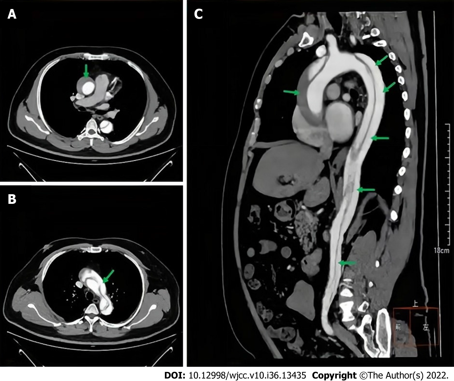
Figure 1 Enhanced computed tomography scan of the patient's aorta.
A: Transverse computed tomography (CT) scan of the ascending aorta; B: Cross-sectional CT images of aortic arch; C: Sagittal CT scan of the aorta. The green arrows indicate the aortic dissection.
- Citation: Yang JH, Wang S, Gan YX, Feng XY, Niu BL. Short-term prone positioning for severe acute respiratory distress syndrome after cardiopulmonary bypass: A case report and literature review. World J Clin Cases 2022; 10(36): 13435-13442
- URL: https://www.wjgnet.com/2307-8960/full/v10/i36/13435.htm
- DOI: https://dx.doi.org/10.12998/wjcc.v10.i36.13435


