Copyright
©The Author(s) 2022.
World J Clin Cases. Dec 26, 2022; 10(36): 13381-13387
Published online Dec 26, 2022. doi: 10.12998/wjcc.v10.i36.13381
Published online Dec 26, 2022. doi: 10.12998/wjcc.v10.i36.13381
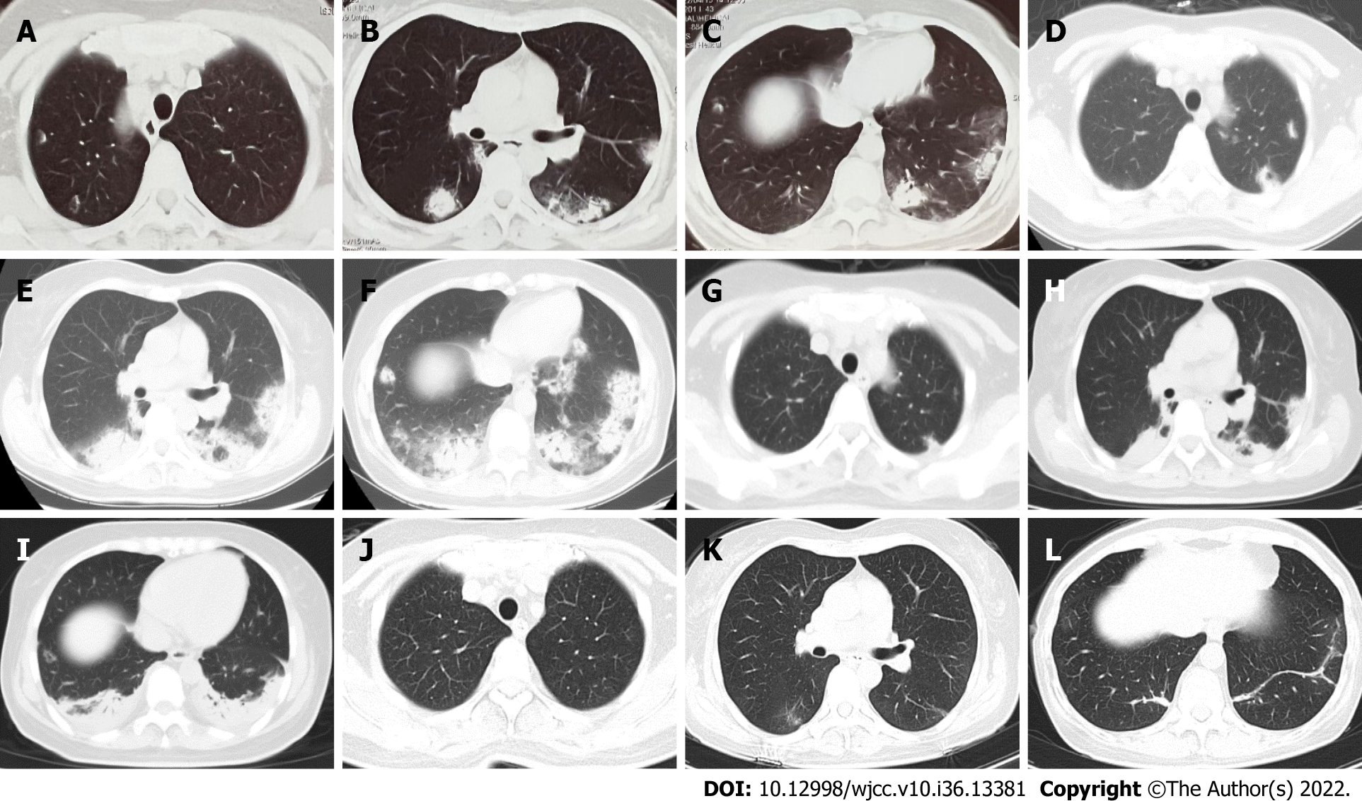
Figure 1 Chest imaging changes.
A–C: Chest computed tomography (CT) at the local hospital suggesting multiple exudative opacities in both lungs; D–F: Chest CT on admission revealed patchy, diffuse alveolar opacities and consolidation with bilateral and peripheral distributions after 9 d of anti-infective treatment in the local hospital; G–I: Chest CT shows that the lung opacities have decreased significantly after 3 d of steroid treatment; J–L: Chest CT shows that the lung opacities have almost been entirely absorbed after 1 mo of steroid treatment.
- Citation: Liu WJ, Zhou S, Li YX. Two methods of lung biopsy for histological confirmation of acute fibrinous and organizing pneumonia: A case report. World J Clin Cases 2022; 10(36): 13381-13387
- URL: https://www.wjgnet.com/2307-8960/full/v10/i36/13381.htm
- DOI: https://dx.doi.org/10.12998/wjcc.v10.i36.13381


