Copyright
©The Author(s) 2022.
World J Clin Cases. Dec 26, 2022; 10(36): 13364-13372
Published online Dec 26, 2022. doi: 10.12998/wjcc.v10.i36.13364
Published online Dec 26, 2022. doi: 10.12998/wjcc.v10.i36.13364
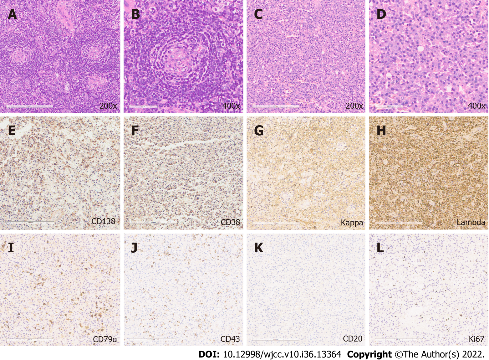
Figure 2 Histopathological and immunohistochemical analysis.
A and B: Hematoxylin and eosin staining revealed angiofollicular lymph node hyperplasia (200 × and 400 ×); C and D: The presence of mature plasma cells (200 × and 400 ×); E-J: Positive immunohistochemical staining for plasma cell markers: CD138, CD38, κ light chains, λ light chains, CD79α, T-cell marker CD43; K: Negative staining for B-cell marker CD20; and L: The Ki-67 positive staining.
- Citation: Zhang YH, He YF, Yue H, Zhang YN, Shi L, Jin B, Dong P. Solitary hyoid plasmacytoma with unicentric Castleman disease: A case report and review of literature. World J Clin Cases 2022; 10(36): 13364-13372
- URL: https://www.wjgnet.com/2307-8960/full/v10/i36/13364.htm
- DOI: https://dx.doi.org/10.12998/wjcc.v10.i36.13364


