Copyright
©The Author(s) 2022.
World J Clin Cases. Dec 26, 2022; 10(36): 13239-13249
Published online Dec 26, 2022. doi: 10.12998/wjcc.v10.i36.13239
Published online Dec 26, 2022. doi: 10.12998/wjcc.v10.i36.13239
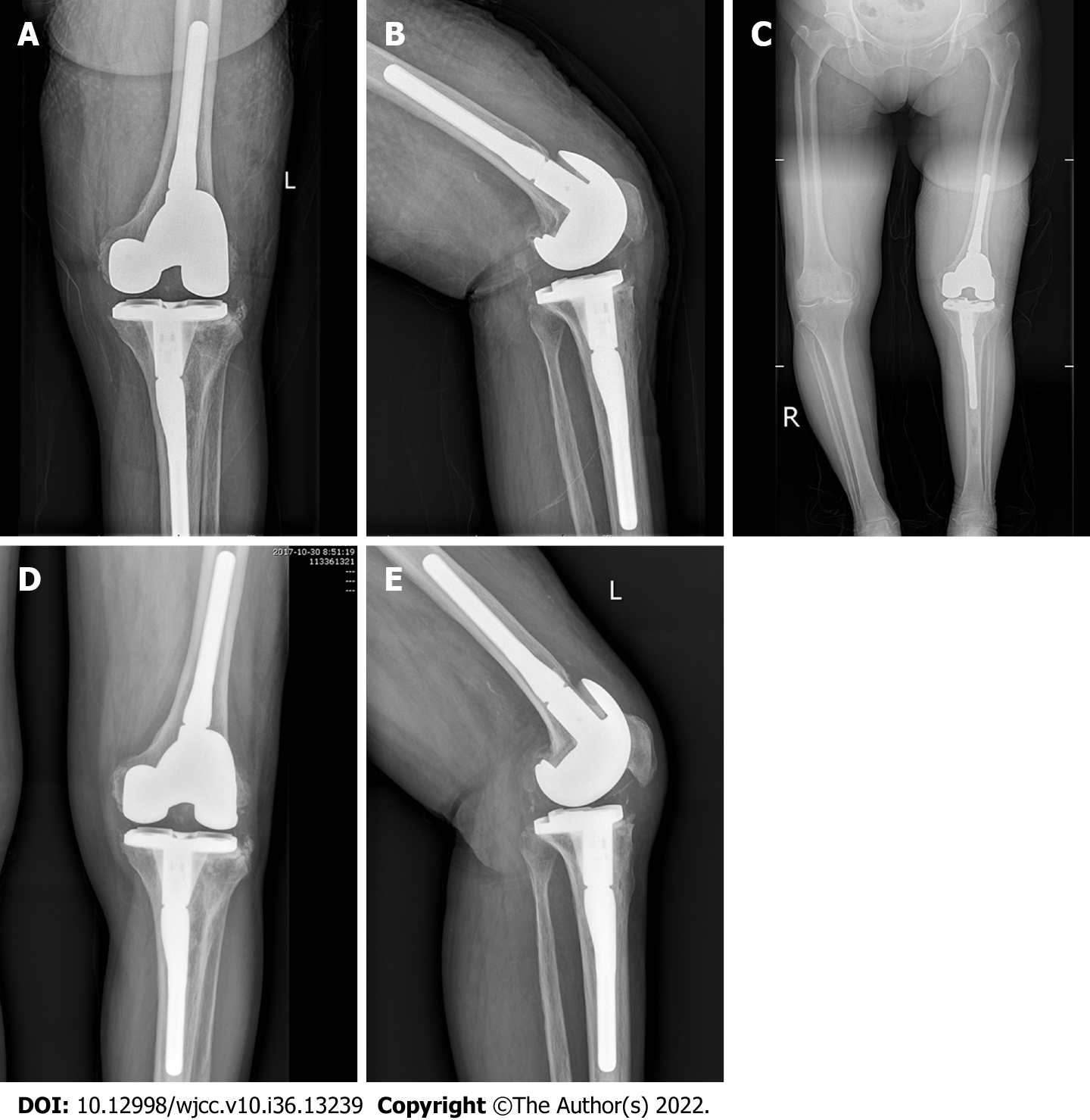
Figure 3 Radiographic image of the left knee of a 78-year-old female patient after phase II revision.
A and B: Radiographic images of the left knee after revision: Anteroposterior (A) and latera (B); C: Full-length orthotopic position of the lower limbs after revision; D and E: Radiographic images of the left knee 2 yr after revision: Anteroposterior (D) and lateral (E).
- Citation: Qiao YJ, Li F, Zhang LD, Yu XY, Zhang HQ, Yang WB, Song XY, Xu RL, Zhou SH. Analysis of the clinical efficacy of two-stage revision surgery in the treatment of periprosthetic joint infection in the knee: A retrospective study. World J Clin Cases 2022; 10(36): 13239-13249
- URL: https://www.wjgnet.com/2307-8960/full/v10/i36/13239.htm
- DOI: https://dx.doi.org/10.12998/wjcc.v10.i36.13239


