Copyright
©The Author(s) 2022.
World J Clin Cases. Dec 16, 2022; 10(35): 13129-13137
Published online Dec 16, 2022. doi: 10.12998/wjcc.v10.i35.13129
Published online Dec 16, 2022. doi: 10.12998/wjcc.v10.i35.13129
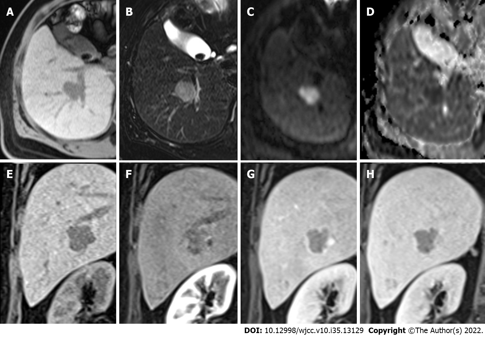
Figure 1 Multiple hepatic nodules at the right hepatic lobe and left hepatic tip are recognized.
A: The representative larger well-defined lesion, 2.5 cm in size, has obvious hypointensity on the fat-suppressed T1WI; B: Hyperintensity on the fat-suppressed T2WI; C: Significant diffusion restriction with hyperintensity on the diffusion-weighted imaging; D: Dark signal on the apparent diffusion coefficient map at the corresponding site; E-H: Early hyperenhancement & rapid washout.
- Citation: Jeng KS, Huang CC, Chung CS, Chang CF. Liver collision tumor of primary hepatocellular carcinoma and neuroendocrine carcinoma: A rare case report. World J Clin Cases 2022; 10(35): 13129-13137
- URL: https://www.wjgnet.com/2307-8960/full/v10/i35/13129.htm
- DOI: https://dx.doi.org/10.12998/wjcc.v10.i35.13129


