Copyright
©The Author(s) 2022.
World J Clin Cases. Dec 16, 2022; 10(35): 13115-13121
Published online Dec 16, 2022. doi: 10.12998/wjcc.v10.i35.13115
Published online Dec 16, 2022. doi: 10.12998/wjcc.v10.i35.13115
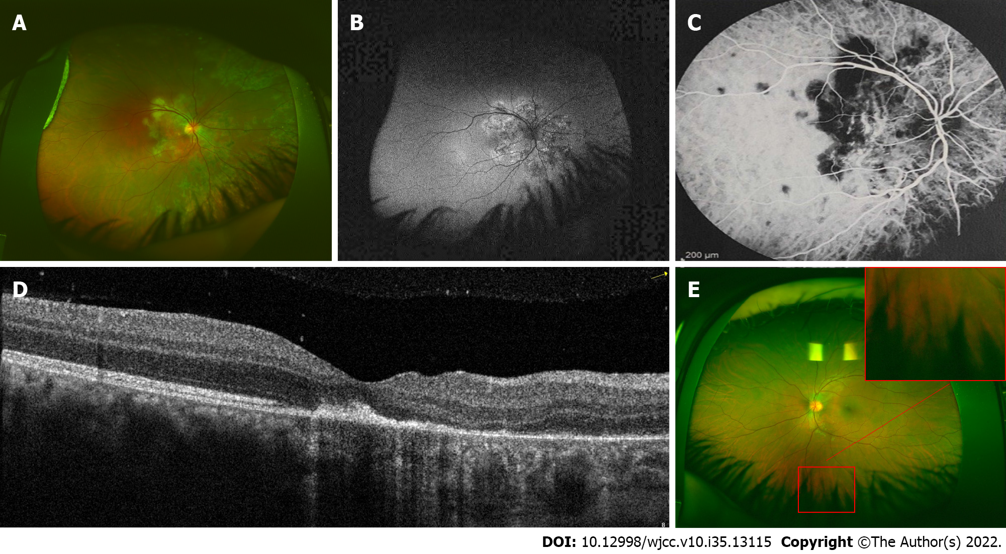
Figure 2 Multimodal imaging of the lesions two weeks after peribulbar injection of triamcinolone.
A: Lesion extension was noted in the right eye, which showed; B: hyperfluorescent on autofluorescence; C: hypofluorescent on indocyanine green angiography; D: Optical coherence tomography of the right eye showed fovea involvement; E: The fundus of the left eye was still unremarkable.
- Citation: Luo L, Chen WB, Zhao MW, Miao H. Systemic combined with intravitreal methotrexate for relentless placoid chorioretinitis: A case report. World J Clin Cases 2022; 10(35): 13115-13121
- URL: https://www.wjgnet.com/2307-8960/full/v10/i35/13115.htm
- DOI: https://dx.doi.org/10.12998/wjcc.v10.i35.13115


