Copyright
©The Author(s) 2022.
World J Clin Cases. Dec 16, 2022; 10(35): 13088-13098
Published online Dec 16, 2022. doi: 10.12998/wjcc.v10.i35.13088
Published online Dec 16, 2022. doi: 10.12998/wjcc.v10.i35.13088
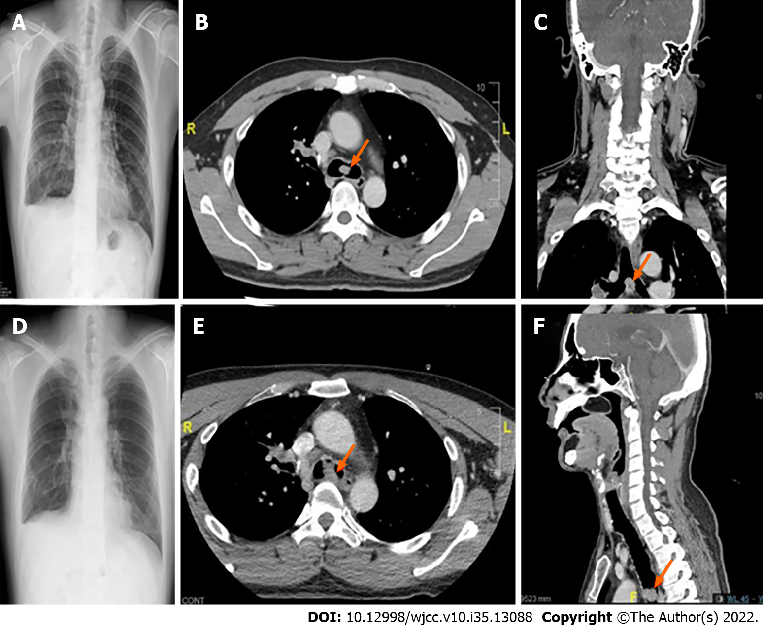
Figure 3 Chest radiography and computed tomography images obtained during previous episodes of general anesthesia.
A-C: Right temporal craniotomy performed at age 40 (October 2019); D-F: oral commissure reposition surgery performed at age 41 (February 2020). These images reveal gradual left lung hyperinflation (A and D), and progressively enlarged carinal masses, approximately 1.2 cm and 2.1 cm in size, respectively. (axial view in B and E; coronal view in C; and sagittal view in F).
- Citation: Liu IL, Chou AH, Chiu CH, Cheng YT, Lin HT. Tracheostomy and venovenous extracorporeal membrane oxygenation for difficult airway patient with carinal melanoma: A case report and literature review. World J Clin Cases 2022; 10(35): 13088-13098
- URL: https://www.wjgnet.com/2307-8960/full/v10/i35/13088.htm
- DOI: https://dx.doi.org/10.12998/wjcc.v10.i35.13088


