Copyright
©The Author(s) 2022.
World J Clin Cases. Dec 16, 2022; 10(35): 13074-13080
Published online Dec 16, 2022. doi: 10.12998/wjcc.v10.i35.13074
Published online Dec 16, 2022. doi: 10.12998/wjcc.v10.i35.13074
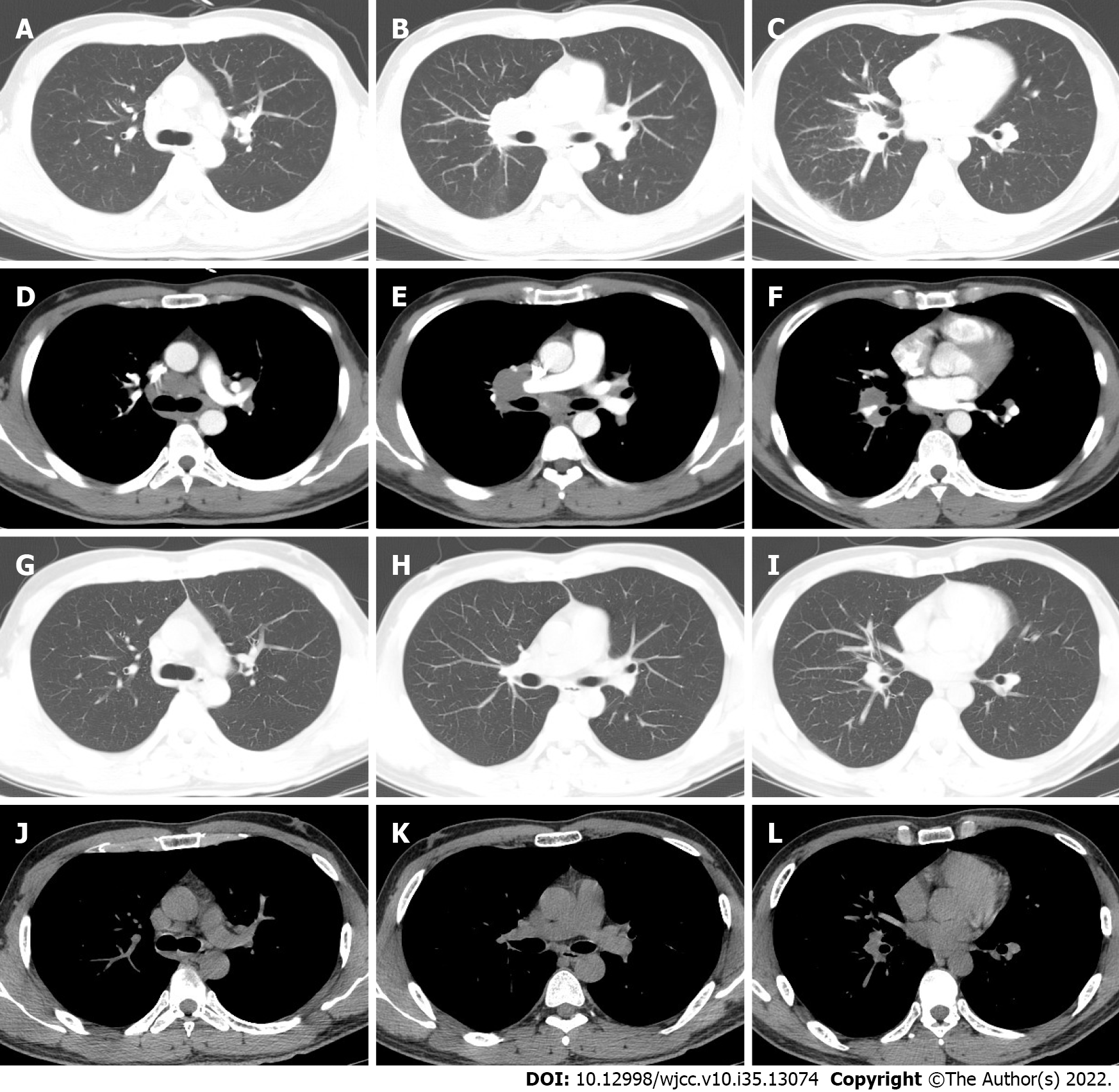
Figure 2 Chest computed tomography.
A-F: On admission, the patient’s chest computed tomography (CT) revealed multiple pulmonary nodules and mediastinal and hilar lymphadenopathy enlargement; G-L: The chest CT reexamination after six months of drug withdrawal revealed a resolution of the pulmonary nodules and a reduction in the size of the mediastinal and hilar lymphadenopathy.
- Citation: Hu YQ, Lv CY, Cui A. Pulmonary sarcoidosis: A novel sequelae of drug reaction with eosinophilia and systemic symptoms: A case report. World J Clin Cases 2022; 10(35): 13074-13080
- URL: https://www.wjgnet.com/2307-8960/full/v10/i35/13074.htm
- DOI: https://dx.doi.org/10.12998/wjcc.v10.i35.13074


