Copyright
©The Author(s) 2022.
World J Clin Cases. Dec 16, 2022; 10(35): 13044-13051
Published online Dec 16, 2022. doi: 10.12998/wjcc.v10.i35.13044
Published online Dec 16, 2022. doi: 10.12998/wjcc.v10.i35.13044
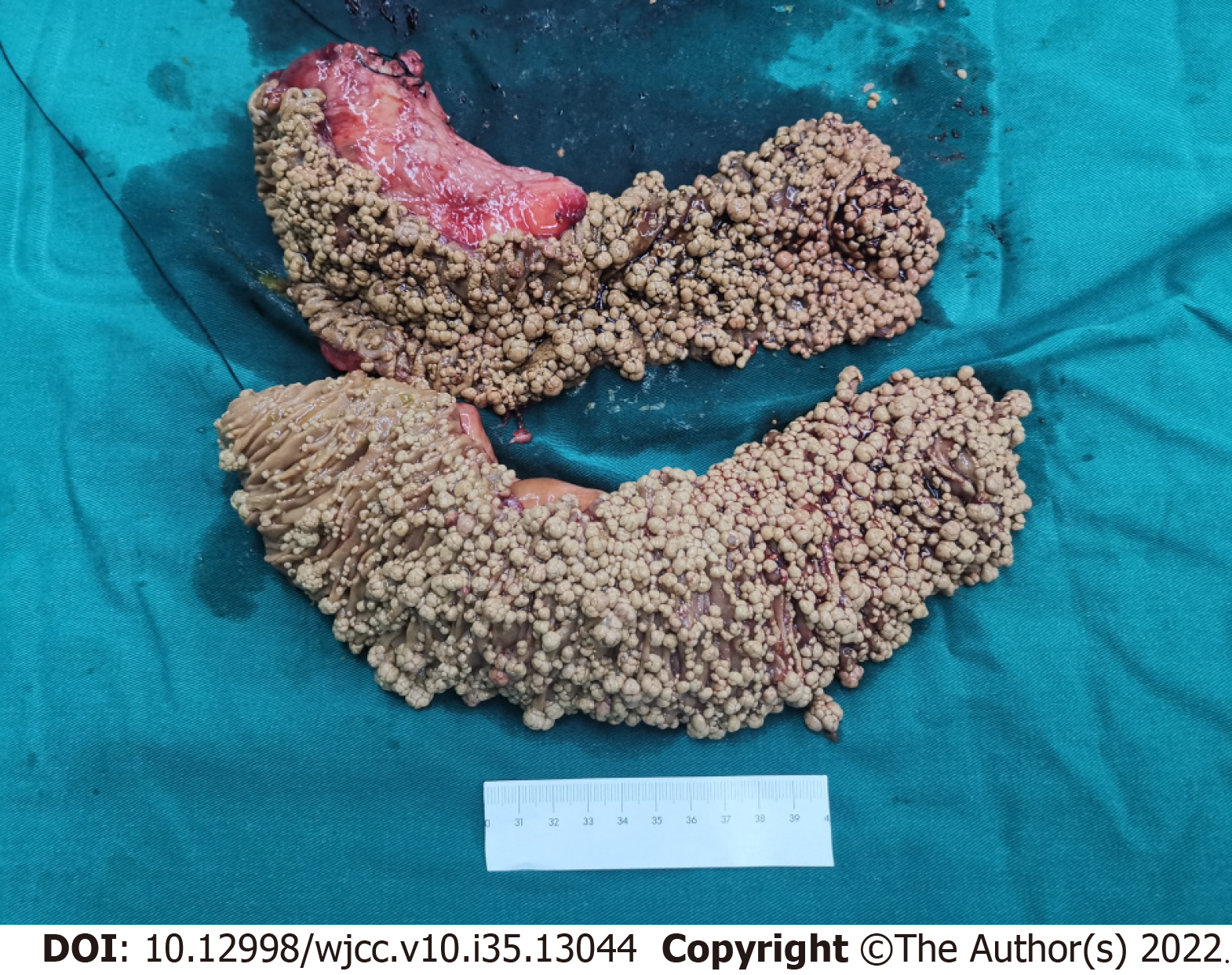
Figure 4 Specimens of the removed pancreatic head and small intestine.
The upper specimen included the pancreatic head (top left) and partial duodenum, and the lower specimen was the proximal jejunum. The mucosal surface of the intestine was covered with grayish-yellow polypoid bulges with various sizes ranging from the needle tip to 2 cm × 1 cm × 1 cm.
- Citation: Chen S, Zhou YC, Si S, Liu HY, Zhang QR, Yin TF, Xie CX, Yao SK, Du SY. Atypical Whipple’s disease with special endoscopic manifestations: A case report. World J Clin Cases 2022; 10(35): 13044-13051
- URL: https://www.wjgnet.com/2307-8960/full/v10/i35/13044.htm
- DOI: https://dx.doi.org/10.12998/wjcc.v10.i35.13044


