Copyright
©The Author(s) 2022.
World J Clin Cases. Dec 16, 2022; 10(35): 13044-13051
Published online Dec 16, 2022. doi: 10.12998/wjcc.v10.i35.13044
Published online Dec 16, 2022. doi: 10.12998/wjcc.v10.i35.13044
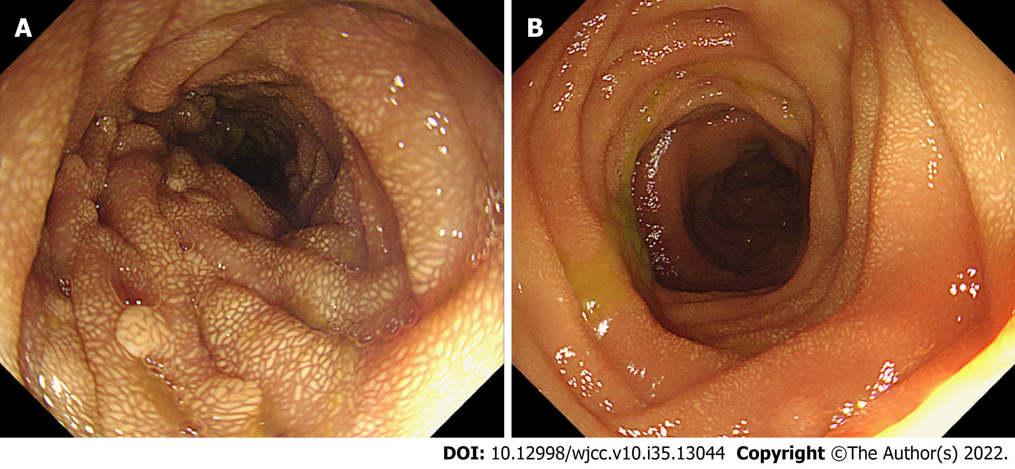
Figure 3 Endoscopic images of the jejunum during the operation.
A: Proximal jejunum; B: Distal jejunum. Multiple polyps were observed in the proximal jejunum and gradually disappeared in the distal jejunum. Whitish-yellow plaque-like changes were diffusely distributed both in the proximal and distal jejunum.
- Citation: Chen S, Zhou YC, Si S, Liu HY, Zhang QR, Yin TF, Xie CX, Yao SK, Du SY. Atypical Whipple’s disease with special endoscopic manifestations: A case report. World J Clin Cases 2022; 10(35): 13044-13051
- URL: https://www.wjgnet.com/2307-8960/full/v10/i35/13044.htm
- DOI: https://dx.doi.org/10.12998/wjcc.v10.i35.13044


