Copyright
©The Author(s) 2022.
World J Clin Cases. Dec 16, 2022; 10(35): 12980-12989
Published online Dec 16, 2022. doi: 10.12998/wjcc.v10.i35.12980
Published online Dec 16, 2022. doi: 10.12998/wjcc.v10.i35.12980
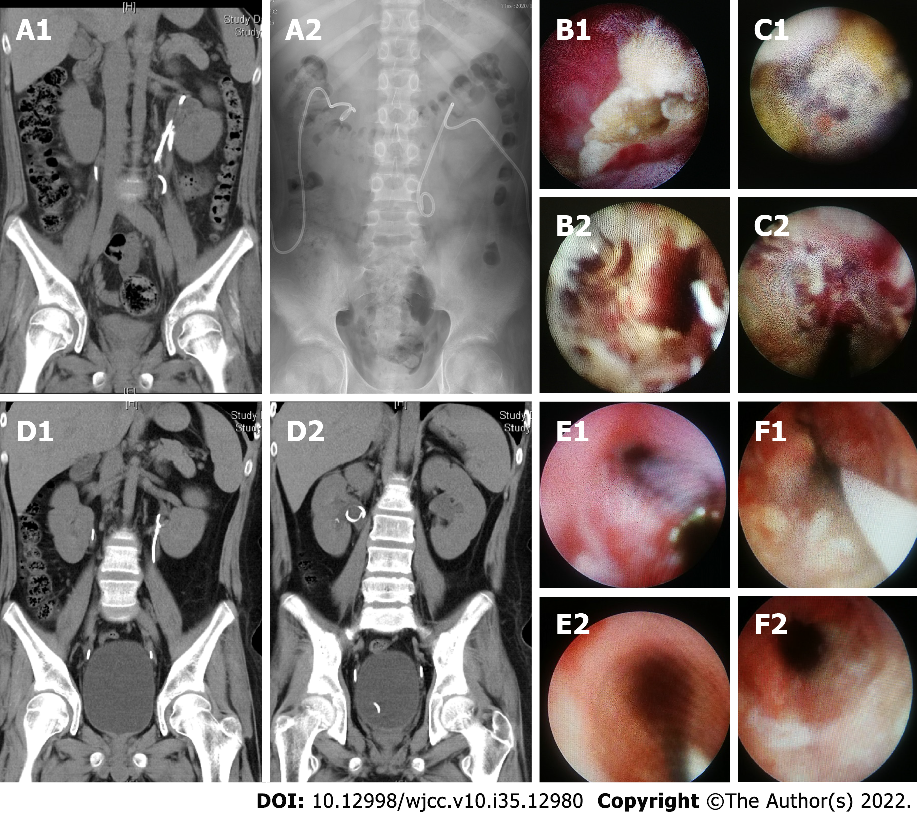
Figure 2 Imaging and endoscopic images of patient 2.
A: Preoperative coronal non-enhanced computed tomography (NCCT) (A1) and KUB (A1) scans showed calcification within the bilateral upper urinary tract and stents of both kidneys; B: The left anterograde lumen before (B1) and after (B2) encrusted tissue removal during the first-stage operation; C: The right retrograde lumen before (C1) and after (C2) encrusted tissue removal during the first-stage operation; D: NCCT scan performed 3 mo postoperatively showed that the bilateral calcification disappeared and the stents were in good positions; E: Right retrograde lumen stent replacement performed 3 mo postoperatively before (E1) and after (E2) encrusted tissue removal; F: Left retrograde lumen stent replacement performed 3 mo postoperatively before (F1) and after (F2) encrusted tissue removal. NCCT: Non-enhanced computed tomography); KUB: Kidney, ureter, and bladder.
- Citation: Liu YB, Xiao B, Hu WG, Zhang G, Fu M, Li JX. Endoscopic treatment of urothelial encrusted pyelo-ureteritis disease: A case series. World J Clin Cases 2022; 10(35): 12980-12989
- URL: https://www.wjgnet.com/2307-8960/full/v10/i35/12980.htm
- DOI: https://dx.doi.org/10.12998/wjcc.v10.i35.12980


