Copyright
©The Author(s) 2022.
World J Clin Cases. Dec 6, 2022; 10(34): 12742-12749
Published online Dec 6, 2022. doi: 10.12998/wjcc.v10.i34.12742
Published online Dec 6, 2022. doi: 10.12998/wjcc.v10.i34.12742
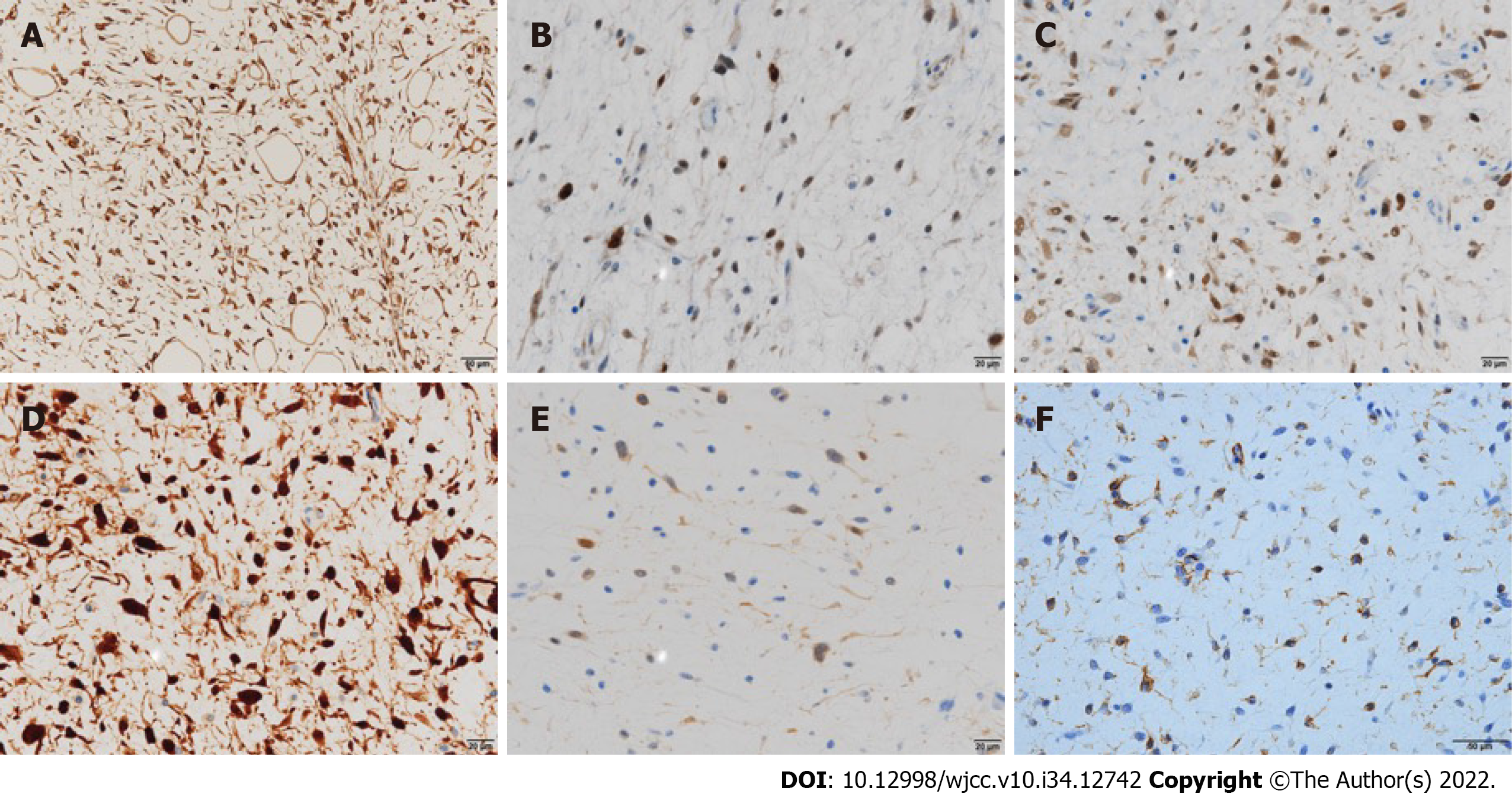
Figure 4 Immunohistochemical examination of the tumor tissue.
Immunohistochemically, the tumor cells were positive for A: vimentin (original magnification: 400 ×; scale bar: 50 µm); B: MDM2 (original magnification: 400 ×; scale bar: 20 µm); C: CDK4 (original magnification: 400 ×; scale bar: 20 µm); and D: p16 (original magnification: 400 ×; scale bar: 20 µm) and focally positive for E: S-100 (original magnification: 400 ×; scale bar: 20 µm) and F: CD34 (original magnification: 400 ×; scale bar: 50 µm).
- Citation: Kugimoto T, Yamagata Y, Ohsako T, Hirai H, Nishii N, Kayamori K, Ikeda T, Harada H. Massive low-grade myxoid liposarcoma of the floor of the mouth: A case report and review of literature. World J Clin Cases 2022; 10(34): 12742-12749
- URL: https://www.wjgnet.com/2307-8960/full/v10/i34/12742.htm
- DOI: https://dx.doi.org/10.12998/wjcc.v10.i34.12742


