Copyright
©The Author(s) 2022.
World J Clin Cases. Dec 6, 2022; 10(34): 12742-12749
Published online Dec 6, 2022. doi: 10.12998/wjcc.v10.i34.12742
Published online Dec 6, 2022. doi: 10.12998/wjcc.v10.i34.12742
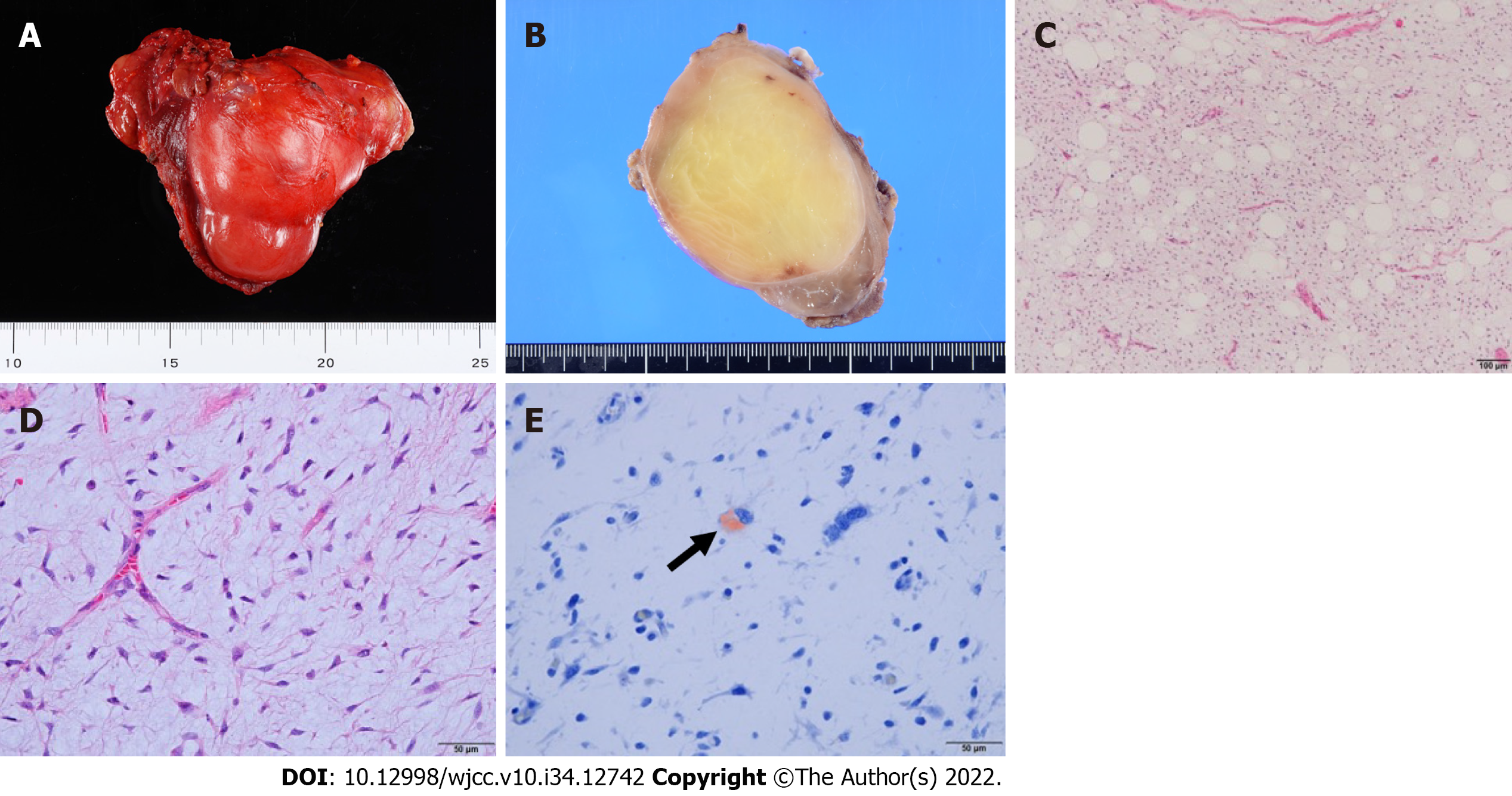
Figure 3 Macroscopic and histological findings of the tumor tissue.
A: The resected specimen was an 8.5 cm × 6.5 cm × 5.5 cm encapsulated tumor; B: Macroscopic findings indicated that the cut surface of the resected tumor was yellowish and greasy; C: Histologically, the tumor comprised uniform primitive mesenchymal cells in an abundant myxoid stroma with delicate, branching, chicken-wire capillary vasculature (original magnification: 100 ×; scale bar: 100 µm); D: Most areasof the lesion exhibited low cellularity. Although round to oval nonlipogenic cells were dominant (original magnification: 400 ×; scale bar: 50 µm); E: Small amounts of Oil Red O-positive lipoblasts were also detected (original magnification: 400 ×; scale bar: 50 µm).
- Citation: Kugimoto T, Yamagata Y, Ohsako T, Hirai H, Nishii N, Kayamori K, Ikeda T, Harada H. Massive low-grade myxoid liposarcoma of the floor of the mouth: A case report and review of literature. World J Clin Cases 2022; 10(34): 12742-12749
- URL: https://www.wjgnet.com/2307-8960/full/v10/i34/12742.htm
- DOI: https://dx.doi.org/10.12998/wjcc.v10.i34.12742


