Copyright
©The Author(s) 2022.
World J Clin Cases. Dec 6, 2022; 10(34): 12742-12749
Published online Dec 6, 2022. doi: 10.12998/wjcc.v10.i34.12742
Published online Dec 6, 2022. doi: 10.12998/wjcc.v10.i34.12742
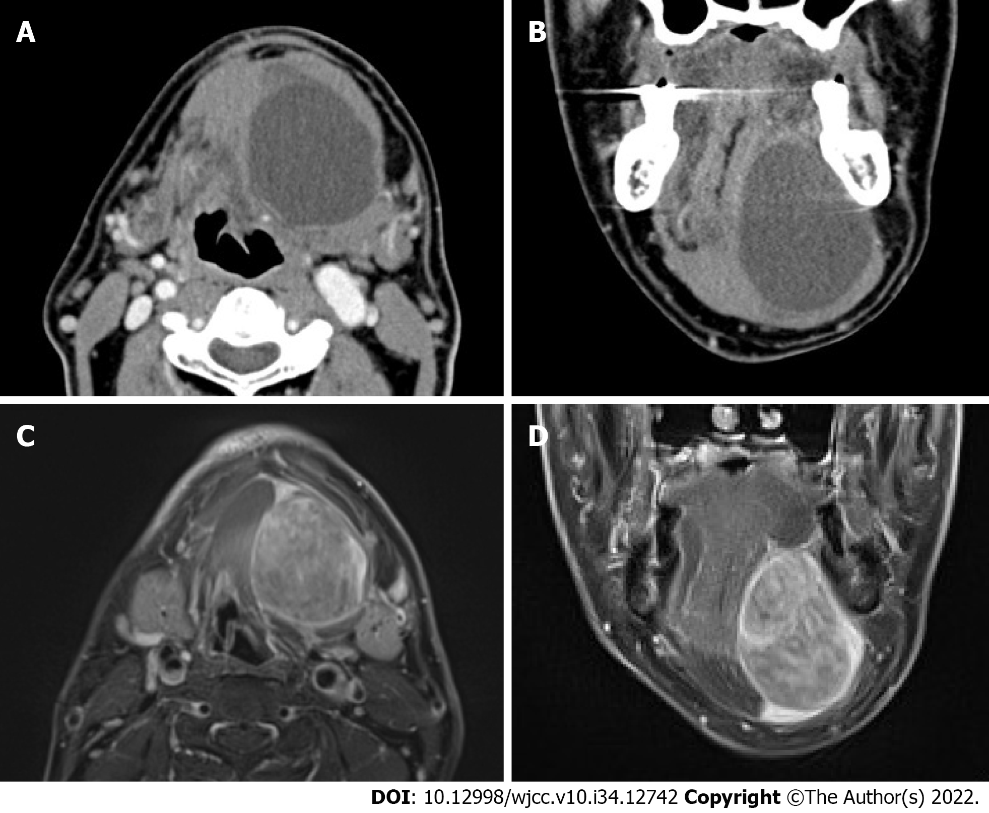
Figure 1 Computed tomography and magnetic resonance imaging findings of myxoid liposarcoma of the floor of the mouth.
A and B: Axial and coronal contrast-enhanced computed tomography findings indicated an unenhanced cystic lesion separating from the surrounding muscle tissue; C and D: Axial and coronal gadolinium-enhanced T1-weighted magnetic resonance imaging showed intensely enhanced tumor lesion. The tumor did not affect the suprahyoid muscles but occupied the sublingual space.
- Citation: Kugimoto T, Yamagata Y, Ohsako T, Hirai H, Nishii N, Kayamori K, Ikeda T, Harada H. Massive low-grade myxoid liposarcoma of the floor of the mouth: A case report and review of literature. World J Clin Cases 2022; 10(34): 12742-12749
- URL: https://www.wjgnet.com/2307-8960/full/v10/i34/12742.htm
- DOI: https://dx.doi.org/10.12998/wjcc.v10.i34.12742


