Copyright
©The Author(s) 2022.
World J Clin Cases. Dec 6, 2022; 10(34): 12654-12664
Published online Dec 6, 2022. doi: 10.12998/wjcc.v10.i34.12654
Published online Dec 6, 2022. doi: 10.12998/wjcc.v10.i34.12654
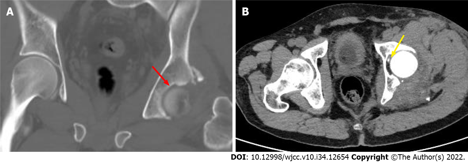
Figure 3 Computed tomography scans.
A, B: The images show an acetabular posterior wall fracture associated with a marginal impaction fracture (red arrow) and an intra-articular loose body (yellow arrow).
- Citation: Xu Y, Lv M, Yu SQ, Liu GP. Closed reduction of hip dislocation associated with ipsilateral lower extremity fractures: A case report and review of the literature. World J Clin Cases 2022; 10(34): 12654-12664
- URL: https://www.wjgnet.com/2307-8960/full/v10/i34/12654.htm
- DOI: https://dx.doi.org/10.12998/wjcc.v10.i34.12654


