Copyright
©The Author(s) 2022.
World J Clin Cases. Dec 6, 2022; 10(34): 12648-12653
Published online Dec 6, 2022. doi: 10.12998/wjcc.v10.i34.12648
Published online Dec 6, 2022. doi: 10.12998/wjcc.v10.i34.12648
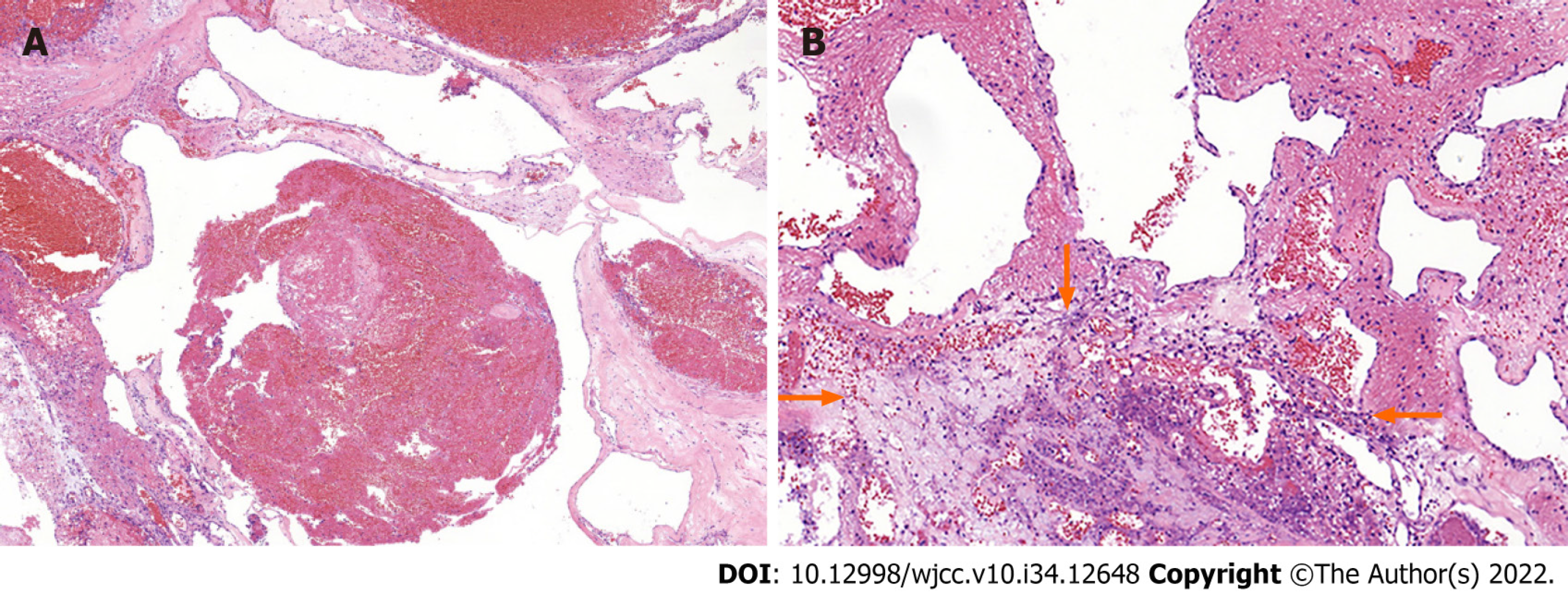
Figure 2 Hematoxylin-eosin staining results (× 200).
The photomicrographs (200 ×) demonstrate blood-filled channels of different diameters with an area of inflammatory necrosis (arrows).
- Citation: Wang GX, Chen YQ, Wang Y, Gao CP. Atypical aggressive vertebral hemangioma of the sacrum with postoperative recurrence: A case report. World J Clin Cases 2022; 10(34): 12648-12653
- URL: https://www.wjgnet.com/2307-8960/full/v10/i34/12648.htm
- DOI: https://dx.doi.org/10.12998/wjcc.v10.i34.12648


