Copyright
©The Author(s) 2022.
World J Clin Cases. Dec 6, 2022; 10(34): 12623-12630
Published online Dec 6, 2022. doi: 10.12998/wjcc.v10.i34.12623
Published online Dec 6, 2022. doi: 10.12998/wjcc.v10.i34.12623
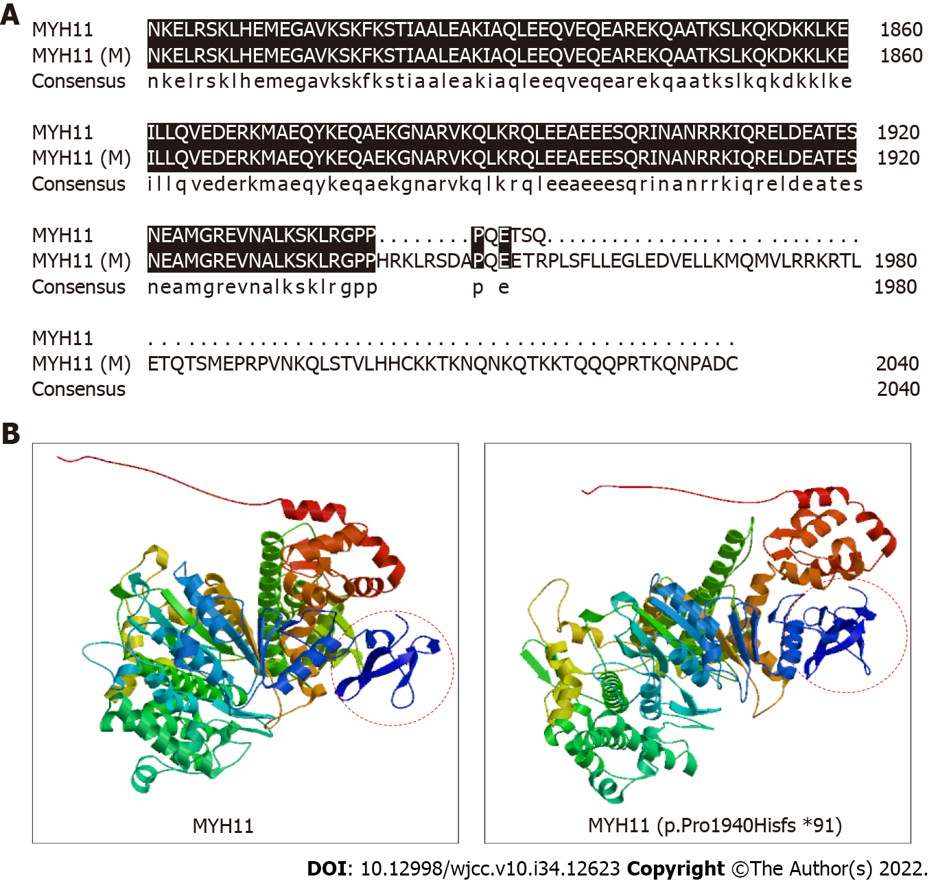
Figure 3 Three-dimensional protein model analysis indicating changes in the structure and conformation of the mutant protein.
A: Alignment of the wild-type MYH11 protein sequence and the MYH11 protein sequence with the p.Pro1940Hisfs*91 mutation from position 1801 using DNAMAN software. Black shading indicates the homologous sequence; B: Three-dimensional structure of the wild-type MYH11 protein and the MYH11 protein sequence with p.Pro1940Hisfs*91 mutation, determined with SWISS-MODEL, a fully automated protein structure homology-modeling server. The mutant protein is 91 amino acids longer than the wild-type MYH11 protein, thus resulting in the change circled in red.
- Citation: Li N, Song YM, Zhang XD, Zhao XS, He XY, Yu LF, Zou DW. Pseudoileus caused by primary visceral myopathy in a Han Chinese patient with a rare MYH11 mutation: A case report. World J Clin Cases 2022; 10(34): 12623-12630
- URL: https://www.wjgnet.com/2307-8960/full/v10/i34/12623.htm
- DOI: https://dx.doi.org/10.12998/wjcc.v10.i34.12623


