Copyright
©The Author(s) 2022.
World J Clin Cases. Dec 6, 2022; 10(34): 12617-12622
Published online Dec 6, 2022. doi: 10.12998/wjcc.v10.i34.12617
Published online Dec 6, 2022. doi: 10.12998/wjcc.v10.i34.12617
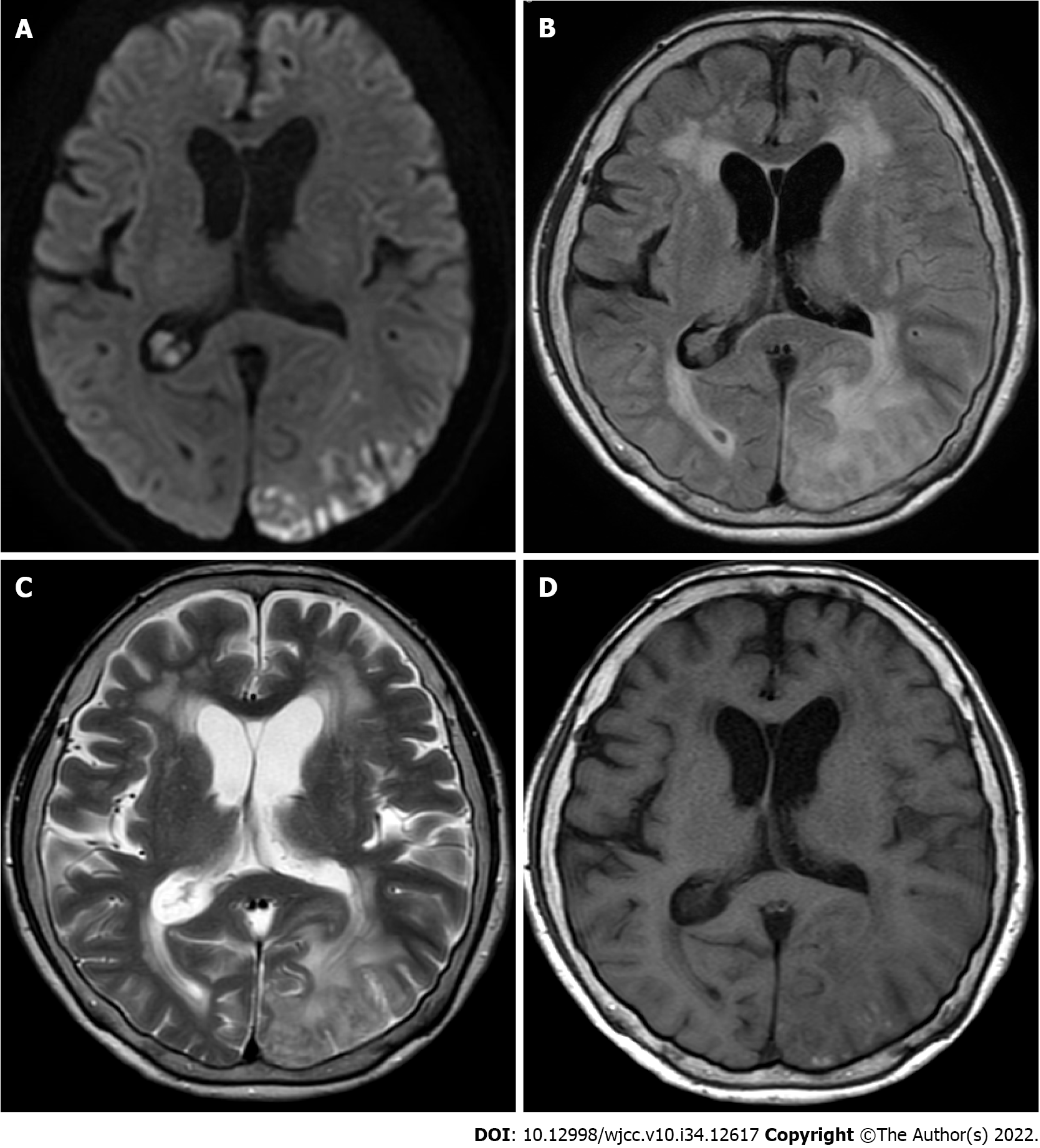
Figure 1 Imaging of the patient’s brain.
A: Diffusion-weighted magnetic resonance imaging showing multiple lesions in the left parieto-occipital lobe that do not match the vascular territories; B: Fluid-attenuated inversion recovery; C: T2-weighted image; D: T1-weighted image.
- Citation: Kizawa M, Iwasaki Y. Amyloid β-related angiitis of the central nervous system occurring after COVID-19 vaccination: A case report. World J Clin Cases 2022; 10(34): 12617-12622
- URL: https://www.wjgnet.com/2307-8960/full/v10/i34/12617.htm
- DOI: https://dx.doi.org/10.12998/wjcc.v10.i34.12617


