Copyright
©The Author(s) 2022.
World J Clin Cases. Nov 26, 2022; 10(33): 12430-12439
Published online Nov 26, 2022. doi: 10.12998/wjcc.v10.i33.12430
Published online Nov 26, 2022. doi: 10.12998/wjcc.v10.i33.12430
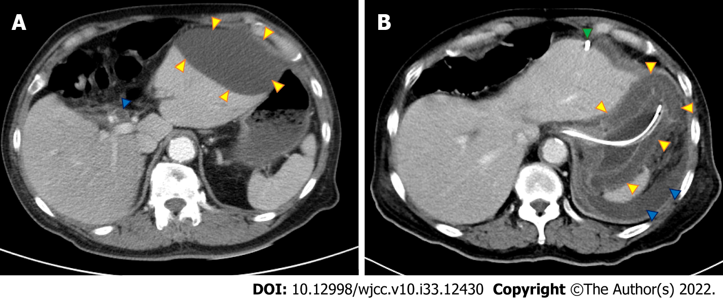
Figure 1 Abdominal computed tomography.
A: Initial abdominal computed tomography (CT) showed a dilated common bile duct (blue arrow) and left subphrenic abscess (yellow arrows); B: Repeat abdominal CT after biloma drainage (green arrow) showed bile leakage (blue arrows) and enlarged wall of the stomach (yellow arrows).
- Citation: Yang KC, Kuo HY, Kang JW. Phlegmonous gastritis after biloma drainage: A case report and review of the literature. World J Clin Cases 2022; 10(33): 12430-12439
- URL: https://www.wjgnet.com/2307-8960/full/v10/i33/12430.htm
- DOI: https://dx.doi.org/10.12998/wjcc.v10.i33.12430


