Copyright
©The Author(s) 2022.
World J Clin Cases. Nov 26, 2022; 10(33): 12380-12387
Published online Nov 26, 2022. doi: 10.12998/wjcc.v10.i33.12380
Published online Nov 26, 2022. doi: 10.12998/wjcc.v10.i33.12380
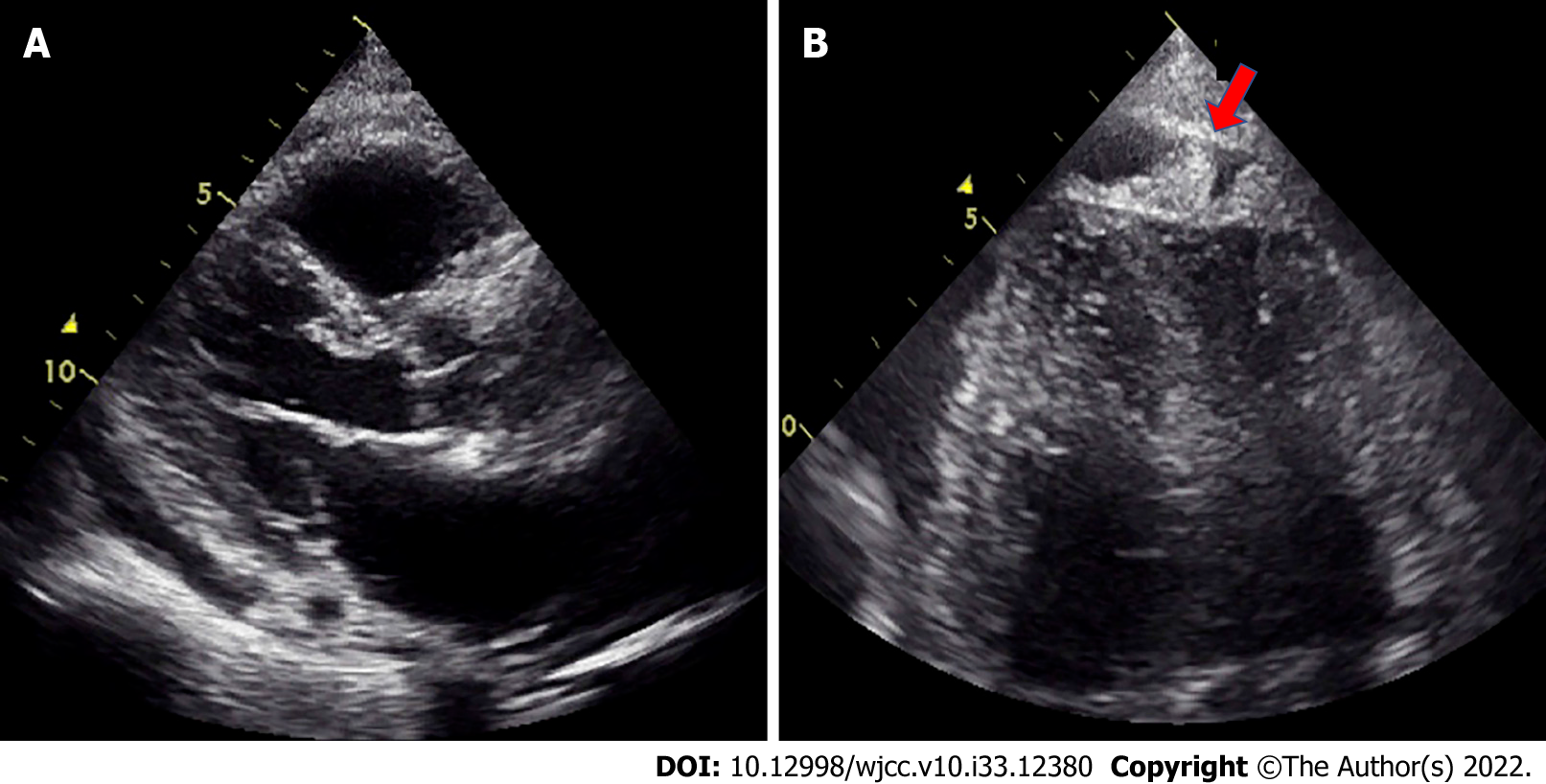
Figure 2 Ultrasound cardiography on day 23.
A: Pericardial effusion was observed, especially in the posterior left ventricle. The degree of pericardial effusion was unchanged; B: Mass-like echogenicity at the apex, which was not present on admission, was evident on the 23rd day (indicated by a red arrow).
- Citation: Oka N, Orita Y, Oshita C, Nakayama H, Teragawa H. Primary malignant pericardial mesothelioma with difficult antemortem diagnosis: A case report. World J Clin Cases 2022; 10(33): 12380-12387
- URL: https://www.wjgnet.com/2307-8960/full/v10/i33/12380.htm
- DOI: https://dx.doi.org/10.12998/wjcc.v10.i33.12380


