Copyright
©The Author(s) 2022.
World J Clin Cases. Nov 26, 2022; 10(33): 12380-12387
Published online Nov 26, 2022. doi: 10.12998/wjcc.v10.i33.12380
Published online Nov 26, 2022. doi: 10.12998/wjcc.v10.i33.12380
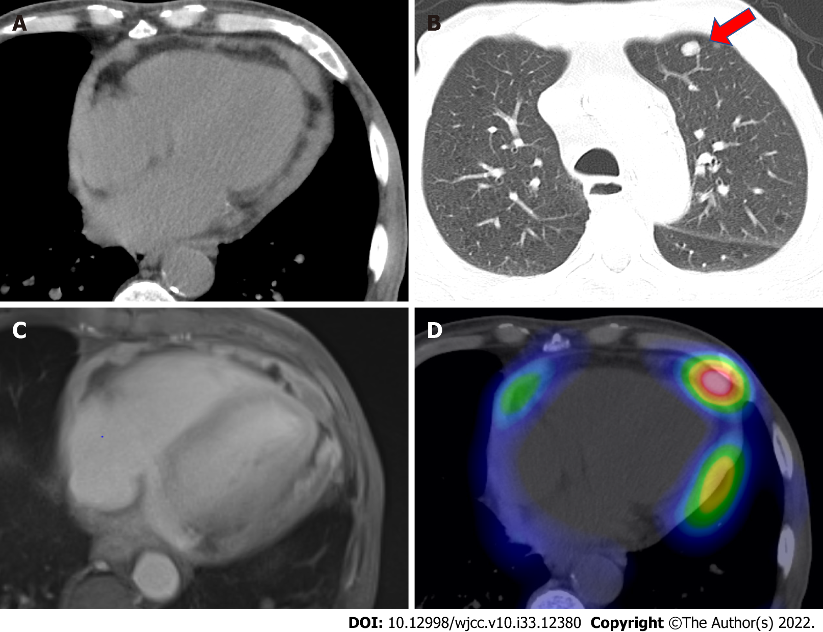
Figure 1 Imaging examinations of the present case.
A: Computed tomography showed cardiac enlargement and high-density pericardial effusion; B: Computed tomography also showed multiple lung metastases (indicated by a red arrow); C: Gadolinium contrast-enhanced T1-weighted images showed thick staining inside and outside the pericardium and a small amount of pericardial fluid; D: Gallium scintigraphy showed several accumulations in the pericardial cavity.
- Citation: Oka N, Orita Y, Oshita C, Nakayama H, Teragawa H. Primary malignant pericardial mesothelioma with difficult antemortem diagnosis: A case report. World J Clin Cases 2022; 10(33): 12380-12387
- URL: https://www.wjgnet.com/2307-8960/full/v10/i33/12380.htm
- DOI: https://dx.doi.org/10.12998/wjcc.v10.i33.12380


