Copyright
©The Author(s) 2022.
World J Clin Cases. Nov 26, 2022; 10(33): 12358-12364
Published online Nov 26, 2022. doi: 10.12998/wjcc.v10.i33.12358
Published online Nov 26, 2022. doi: 10.12998/wjcc.v10.i33.12358
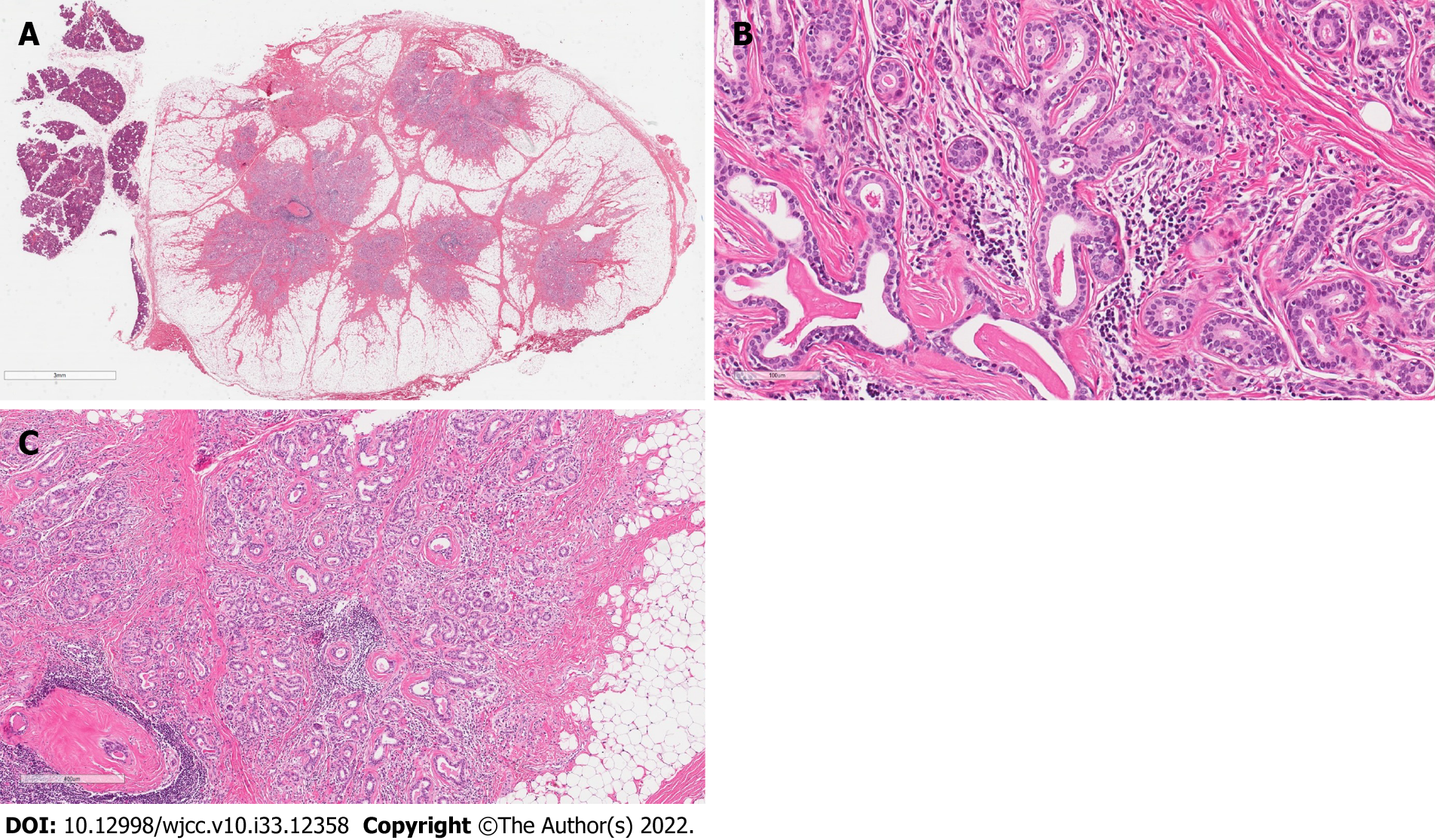
Figure 3 Pathological morphology of the tumor.
A: Beside of normal salivary gland (lobules on the left side of the picture) there was a nodule of the regular border, encapsulated by a thin layer of fibrous tissue. The lesion was composed of numerous small tubules surrounded by a large amount of adipose tissue; B: The tubules were surrounded by a collagenous membrane and consisted of two cell layers: cuboidal ductal and myoepithelial cells; C: There was evidence of fibrosis, periductal hyalinization and focal periductal chronic inflammation.
- Citation: Stankevicius D, Petroska D, Zaleckas L, Kutanovaite O. Hybrid intercalated duct lesion of the parotid: A case report. World J Clin Cases 2022; 10(33): 12358-12364
- URL: https://www.wjgnet.com/2307-8960/full/v10/i33/12358.htm
- DOI: https://dx.doi.org/10.12998/wjcc.v10.i33.12358


