Copyright
©The Author(s) 2022.
World J Clin Cases. Nov 26, 2022; 10(33): 12305-12312
Published online Nov 26, 2022. doi: 10.12998/wjcc.v10.i33.12305
Published online Nov 26, 2022. doi: 10.12998/wjcc.v10.i33.12305
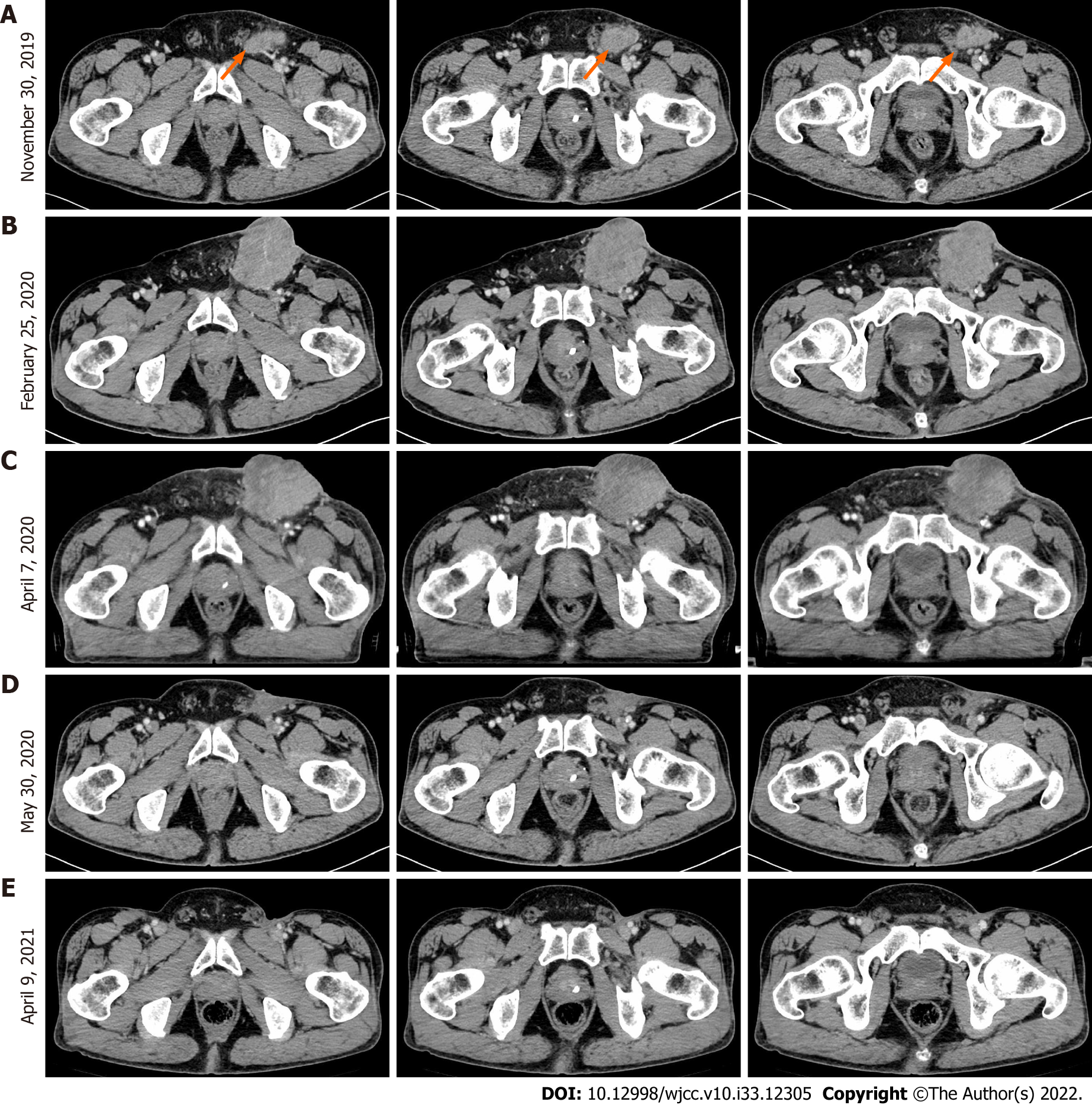
Figure 1 Computed tomography images at different treatment periods.
A: November 30, 2019: Computed tomography (CT) images before epidermal growth factor receptor monoclonal antibody therapy; B: February 25, 2020: CT images before chemotherapy but after epidermal growth factor receptor monoclonal antibody treatment; C: April 7, 2020: CT image of tumor lesions after the first round of chemotherapy alone; D: May 30, 2020: CT image for preoperative evaluation after the third round of immunotherapy combined with chemotherapy; E: April 09, 2021: CT images at the 10-mo follow-up after bilateral inguinal lymph node dissection.
- Citation: Long XY, Zhang S, Tang LS, Li X, Liu JY. Conversion therapy for advanced penile cancer with tislelizumab combined with chemotherapy: A case report and review of literature. World J Clin Cases 2022; 10(33): 12305-12312
- URL: https://www.wjgnet.com/2307-8960/full/v10/i33/12305.htm
- DOI: https://dx.doi.org/10.12998/wjcc.v10.i33.12305


