Copyright
©The Author(s) 2022.
World J Clin Cases. Nov 16, 2022; 10(32): 12036-12044
Published online Nov 16, 2022. doi: 10.12998/wjcc.v10.i32.12036
Published online Nov 16, 2022. doi: 10.12998/wjcc.v10.i32.12036
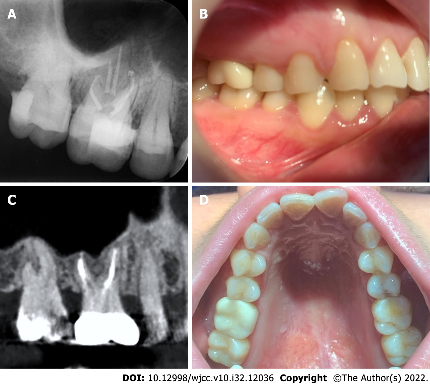
Figure 4 Final crown restoration of the right maxillary first molar.
A: The X-ray at the 1-mo follow-up reveals that the five root canals of the tooth are well obturated; B: Final ceramic crown restoration is performed in the maxillary first molar; C: Radiographic image at the 9-mo follow-up; D: Clinical image at the 9-mo follow-up.
- Citation: Chen K, Ran X, Wang Y. Endodontic treatment of the maxillary first molar with palatal canal variations: A case report and review of literature. World J Clin Cases 2022; 10(32): 12036-12044
- URL: https://www.wjgnet.com/2307-8960/full/v10/i32/12036.htm
- DOI: https://dx.doi.org/10.12998/wjcc.v10.i32.12036


