Copyright
©The Author(s) 2022.
World J Clin Cases. Nov 16, 2022; 10(32): 12036-12044
Published online Nov 16, 2022. doi: 10.12998/wjcc.v10.i32.12036
Published online Nov 16, 2022. doi: 10.12998/wjcc.v10.i32.12036
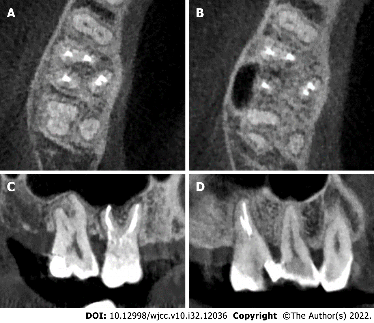
Figure 3 The anatomical structure of the right maxillary first molar was analyzed by cone-beam computed tomography.
A: The cone-beam computed tomography (CBCT) image reveals that the maxillary first molar contains three roots; B: The axial sectional CBCT image demonstrates that the distobuccal (DB) root has one canal, and the mesiobuccal (MB) and palatal roots have two separate canals; C: The sagittal sectional CBCT image reveals that the MB, MB2, and DB canals are well filled; D: The sagittal sectional CBCT image indicates that the mesiopalatal and distopalatal canals in the palatal root are obturated.
- Citation: Chen K, Ran X, Wang Y. Endodontic treatment of the maxillary first molar with palatal canal variations: A case report and review of literature. World J Clin Cases 2022; 10(32): 12036-12044
- URL: https://www.wjgnet.com/2307-8960/full/v10/i32/12036.htm
- DOI: https://dx.doi.org/10.12998/wjcc.v10.i32.12036


