Copyright
©The Author(s) 2022.
World J Clin Cases. Nov 16, 2022; 10(32): 12000-12006
Published online Nov 16, 2022. doi: 10.12998/wjcc.v10.i32.12000
Published online Nov 16, 2022. doi: 10.12998/wjcc.v10.i32.12000
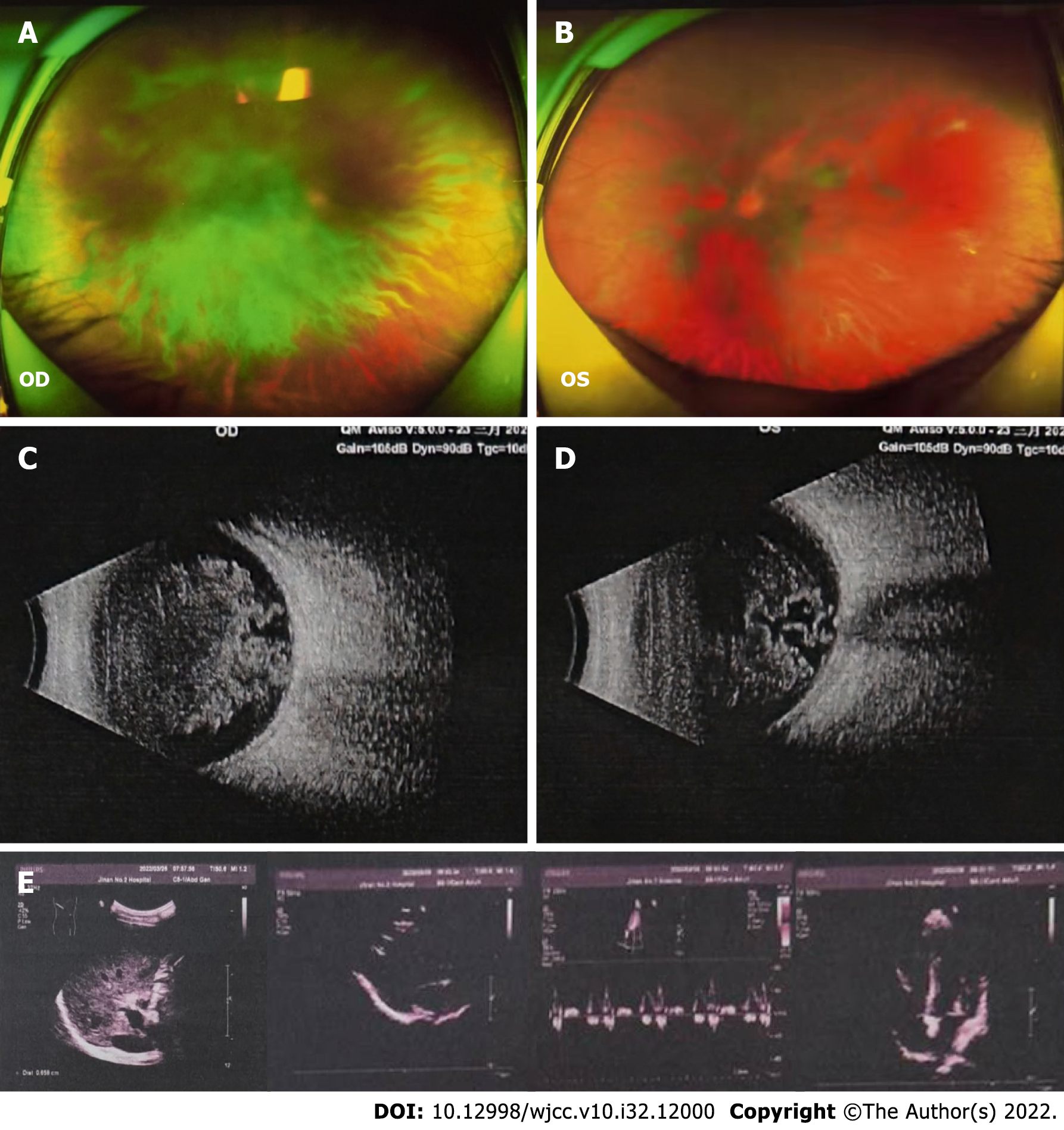
Figure 4 Imaging examinations.
A-E: Fundus color photography (A and B) and ultrasound examination (C and D) showed obvious opacity in bilateral vitreous bodies. There was no obvious abnormality of the heart or abdomen by color ultrasound (E).
- Citation: Tan Y, Tao Y, Sheng YJ, Zhang CM. Vitreous amyloidosis caused by a Lys55Asn variant in transthyretin: A case report. World J Clin Cases 2022; 10(32): 12000-12006
- URL: https://www.wjgnet.com/2307-8960/full/v10/i32/12000.htm
- DOI: https://dx.doi.org/10.12998/wjcc.v10.i32.12000


