Copyright
©The Author(s) 2022.
World J Clin Cases. Nov 16, 2022; 10(32): 12000-12006
Published online Nov 16, 2022. doi: 10.12998/wjcc.v10.i32.12000
Published online Nov 16, 2022. doi: 10.12998/wjcc.v10.i32.12000
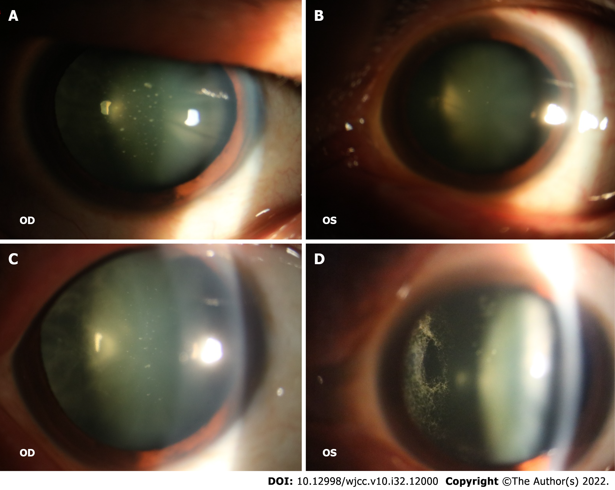
Figure 1 Physical examination.
A and B: White spots on the posterior surface of the lens in both eyes, which were similar to foot plates; C and D: Fundus examination revealed vitreous opacity in both eyes, with a noticeable spider-web-like white cord.
- Citation: Tan Y, Tao Y, Sheng YJ, Zhang CM. Vitreous amyloidosis caused by a Lys55Asn variant in transthyretin: A case report. World J Clin Cases 2022; 10(32): 12000-12006
- URL: https://www.wjgnet.com/2307-8960/full/v10/i32/12000.htm
- DOI: https://dx.doi.org/10.12998/wjcc.v10.i32.12000


