Copyright
©The Author(s) 2022.
World J Clin Cases. Nov 16, 2022; 10(32): 11980-11986
Published online Nov 16, 2022. doi: 10.12998/wjcc.v10.i32.11980
Published online Nov 16, 2022. doi: 10.12998/wjcc.v10.i32.11980
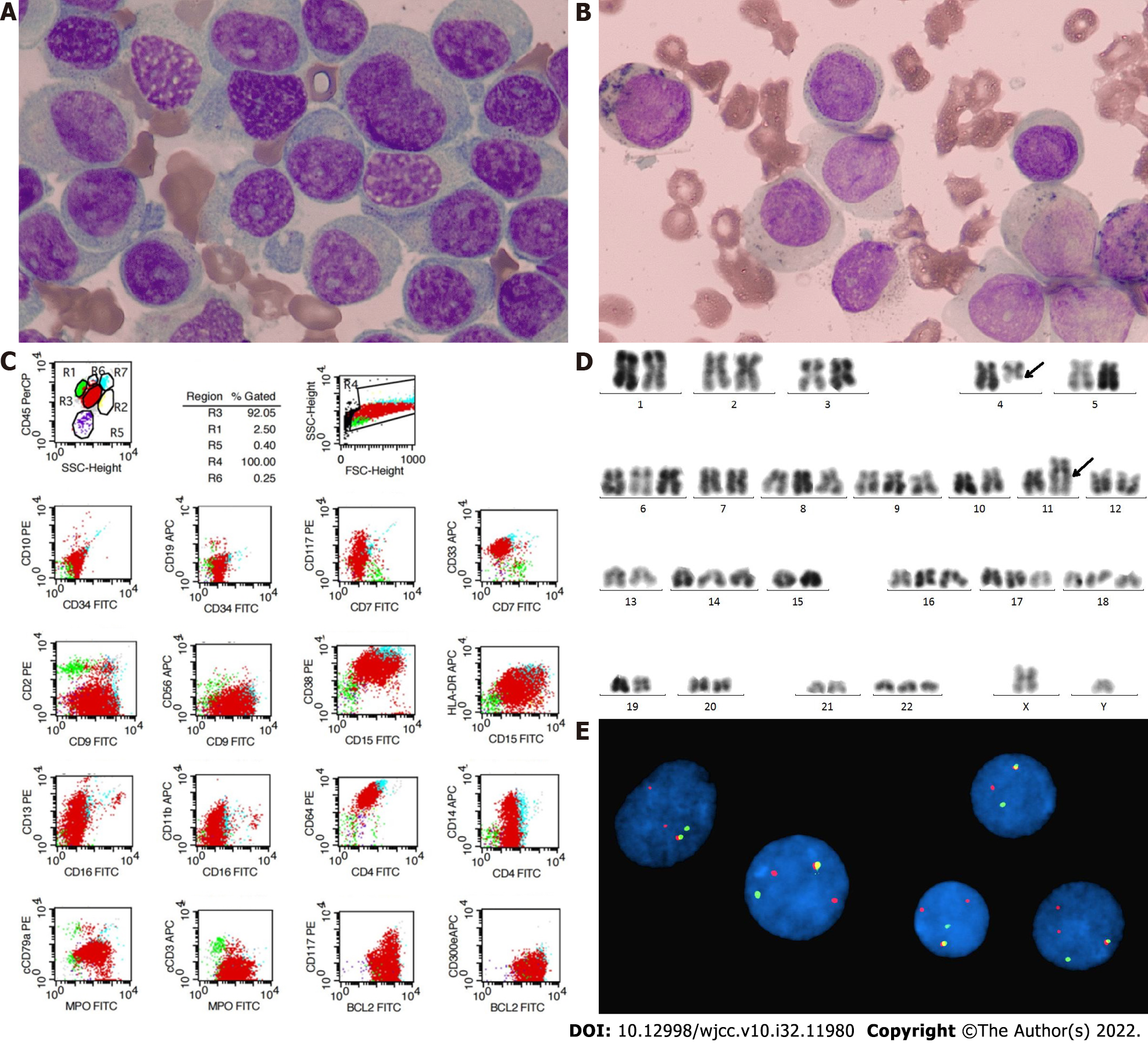
Figure 1 Bone marrow examination at diagnosis.
A: Bone marrow (BM) smear showed large and irregular cells, with rich and dusty blue cytoplasm and a few azurophilic granules; chromatin was rough and loose, light purple red, and nucleoli were not clear; B: Peroxidase staining was weak positive; C: Flow cytometry showed that 91.87% myeloid cells in BM were malignant clones, expressing CD33+, CD9+, CD38+, CD15+, CD64+, cMPO+, BCL2+, HLA-DR+/-, CD13+/-, and CD14+/-; D: R-banded cytogenetic test showed a hyperdiploid karyotype with addition of chromosomes 6, 8, 9, 14, 16, 17, 18 and 22 and t(4;11)(q21;q23) balanced translocation; E: Fluorescence in situ hybridization showed MLL break-apart probe (Y), 1R1G1Y signal and 2R1G1Y atypical signal.
- Citation: Zhang MY, Zhao Y, Zhang JH. t(4;11) translocation in hyperdiploid de novo adult acute myeloid leukemia: A case report. World J Clin Cases 2022; 10(32): 11980-11986
- URL: https://www.wjgnet.com/2307-8960/full/v10/i32/11980.htm
- DOI: https://dx.doi.org/10.12998/wjcc.v10.i32.11980


