Copyright
©The Author(s) 2022.
World J Clin Cases. Nov 16, 2022; 10(32): 11853-11860
Published online Nov 16, 2022. doi: 10.12998/wjcc.v10.i32.11853
Published online Nov 16, 2022. doi: 10.12998/wjcc.v10.i32.11853
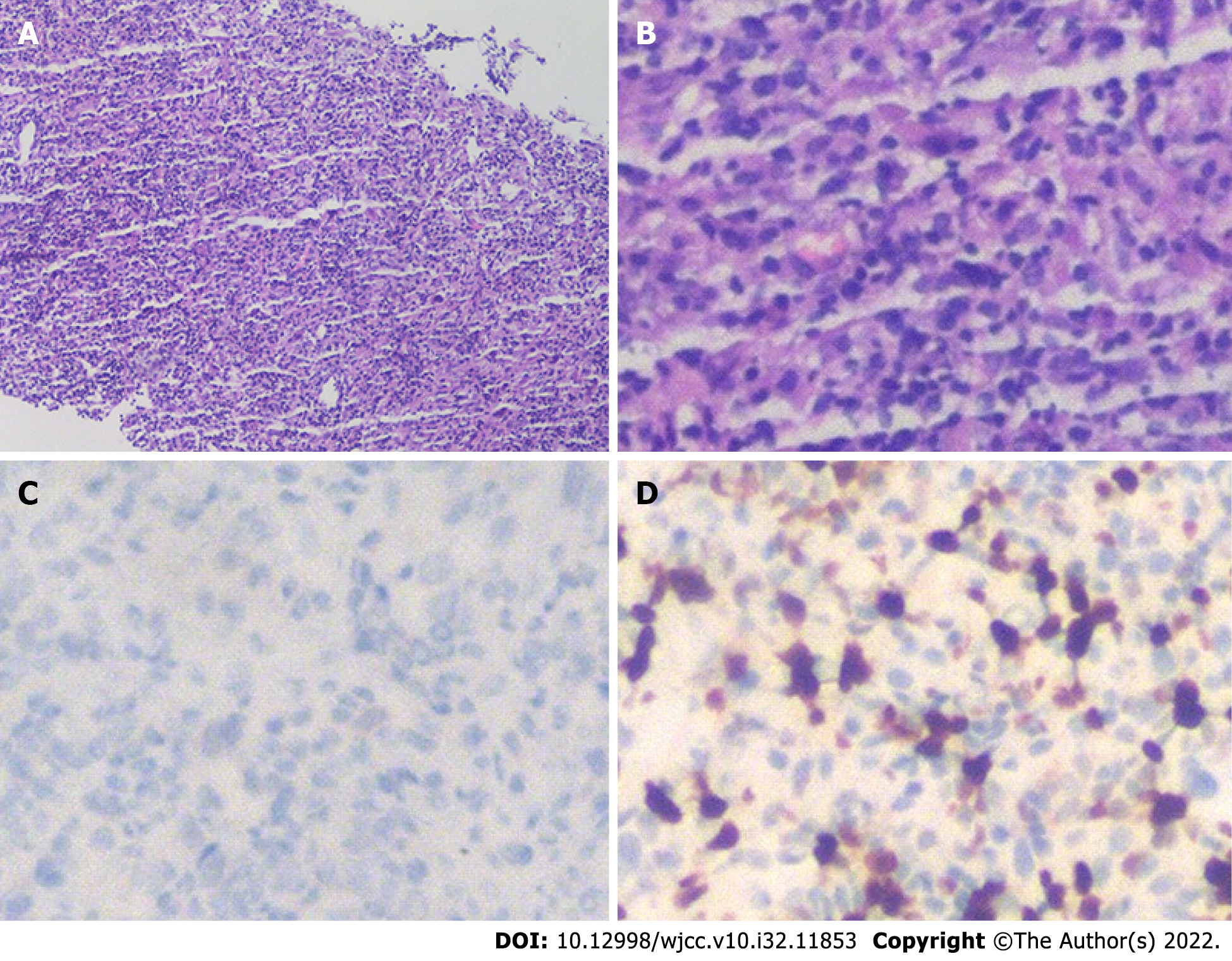
Figure 2 Microscopic examination results of liver biopsy under ultrasound guidance.
A: Routine HE staining showed that there was no obvious monoclonal proliferation of cells (100×), and immunohistochemistry showed that the mass was composed of lymphocytes, myofibroblasts, spindle cells and plasma cells; B: At higher magnification (400×), HE staining was more obvious; C and D: ALK-D5F3 (C) and Ki-67 (D) immunohistochemistry of spindle cells was positive (400×). HE: Hematoxylin and eosin.
- Citation: Li YY, Zang JF, Zhang C. Laparoscopic treatment of inflammatory myofibroblastic tumor in liver: A case report. World J Clin Cases 2022; 10(32): 11853-11860
- URL: https://www.wjgnet.com/2307-8960/full/v10/i32/11853.htm
- DOI: https://dx.doi.org/10.12998/wjcc.v10.i32.11853


