Copyright
©The Author(s) 2022.
World J Clin Cases. Nov 6, 2022; 10(31): 11630-11637
Published online Nov 6, 2022. doi: 10.12998/wjcc.v10.i31.11630
Published online Nov 6, 2022. doi: 10.12998/wjcc.v10.i31.11630
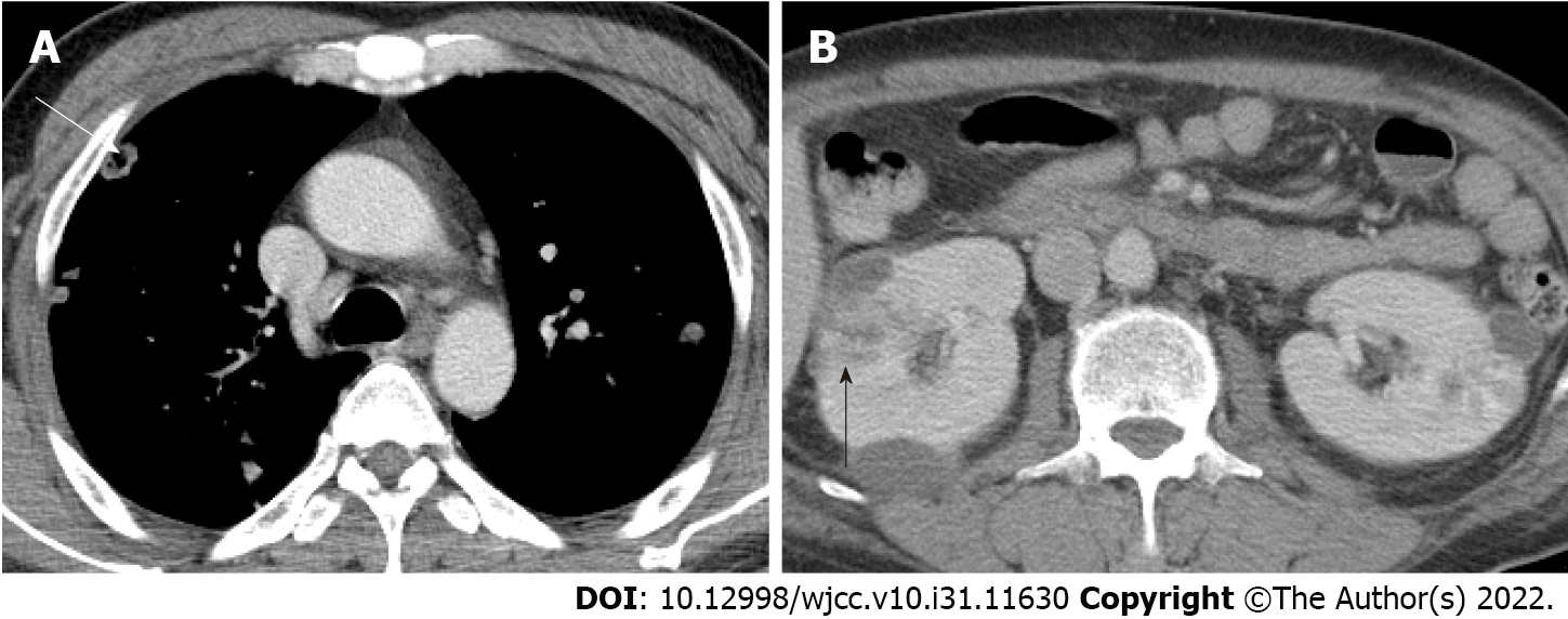
Figure 3 Computed tomographic imaging of internal organ abscesses.
A: Septic emboli on chest computed tomography (white arrow); B: Large renal abscess lesions are observed on abdominopelvic computed tomography (black arrow).
- Citation: Lee DW, Kwak SH, Choi HJ. Secondary craniofacial necrotizing fasciitis from a distant septic emboli: A case report. World J Clin Cases 2022; 10(31): 11630-11637
- URL: https://www.wjgnet.com/2307-8960/full/v10/i31/11630.htm
- DOI: https://dx.doi.org/10.12998/wjcc.v10.i31.11630


