Copyright
©The Author(s) 2022.
World J Clin Cases. Nov 6, 2022; 10(31): 11607-11616
Published online Nov 6, 2022. doi: 10.12998/wjcc.v10.i31.11607
Published online Nov 6, 2022. doi: 10.12998/wjcc.v10.i31.11607
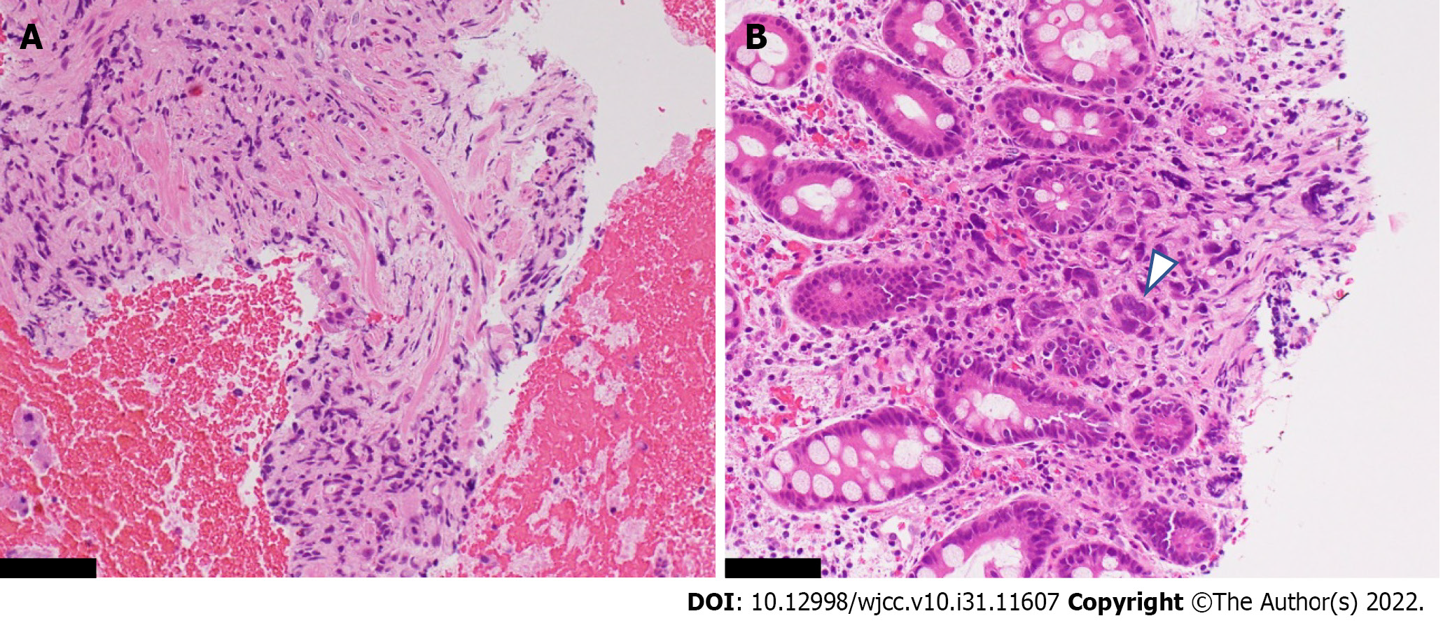
Figure 4 Histopathological specimen obtained by an endoscopic ultrasonography-guided fine-needle biopsy of the thickened gastric wall.
A: Within the intricate muscularis propria, fibroblasts were proliferating, while a few scattered cells suspected of malignancy were seen; B: Poorly differentiated adenocarcinoma cells were seen within the deeper portion of the hyperplastic mucosa (arrowhead). The black scale bar represents 250 μm.
- Citation: Sato R, Matsumoto K, Kanzaki H, Matsumi A, Miyamoto K, Morimoto K, Terasawa H, Fujii Y, Yamazaki T, Uchida D, Tsutsumi K, Horiguchi S, Kato H. Gastric linitis plastica with autoimmune pancreatitis diagnosed by an endoscopic ultrasonography-guided fine-needle biopsy: A case report. World J Clin Cases 2022; 10(31): 11607-11616
- URL: https://www.wjgnet.com/2307-8960/full/v10/i31/11607.htm
- DOI: https://dx.doi.org/10.12998/wjcc.v10.i31.11607


