Copyright
©The Author(s) 2022.
World J Clin Cases. Nov 6, 2022; 10(31): 11607-11616
Published online Nov 6, 2022. doi: 10.12998/wjcc.v10.i31.11607
Published online Nov 6, 2022. doi: 10.12998/wjcc.v10.i31.11607
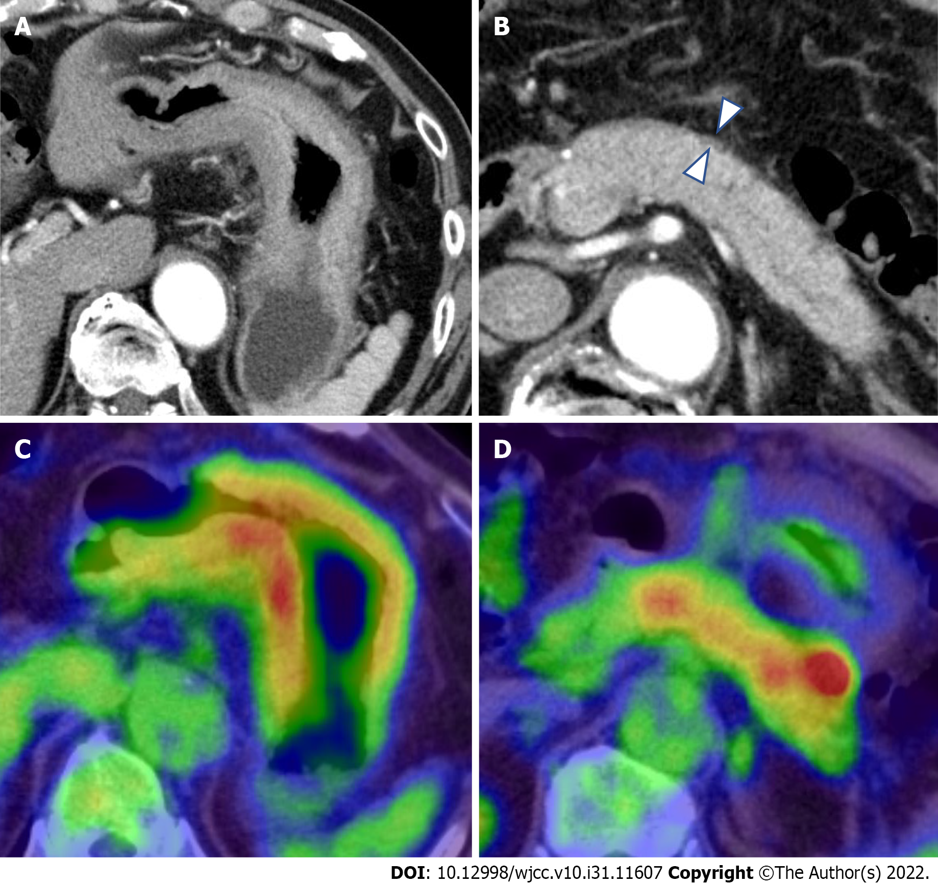
Figure 2 Computed tomography and 18F-Fluorodeoxyglucose-positron emission tomography/computed tomography findings.
A: Contrast-enhanced computed tomography (CT) showed thickening of the wall of the gastric body; B: CT showed the diffuse enlargement of the pancreas and peripancreatic rim (arrowheads); C and D: Fluorodeoxyglucose-positron emission tomography/CT (FDG-PET/CT) showed the accumulation of FDG within both the gastric wall (SUVmax: 19.2) and pancreas.
- Citation: Sato R, Matsumoto K, Kanzaki H, Matsumi A, Miyamoto K, Morimoto K, Terasawa H, Fujii Y, Yamazaki T, Uchida D, Tsutsumi K, Horiguchi S, Kato H. Gastric linitis plastica with autoimmune pancreatitis diagnosed by an endoscopic ultrasonography-guided fine-needle biopsy: A case report. World J Clin Cases 2022; 10(31): 11607-11616
- URL: https://www.wjgnet.com/2307-8960/full/v10/i31/11607.htm
- DOI: https://dx.doi.org/10.12998/wjcc.v10.i31.11607


