Copyright
©The Author(s) 2022.
World J Clin Cases. Nov 6, 2022; 10(31): 11597-11606
Published online Nov 6, 2022. doi: 10.12998/wjcc.v10.i31.11597
Published online Nov 6, 2022. doi: 10.12998/wjcc.v10.i31.11597
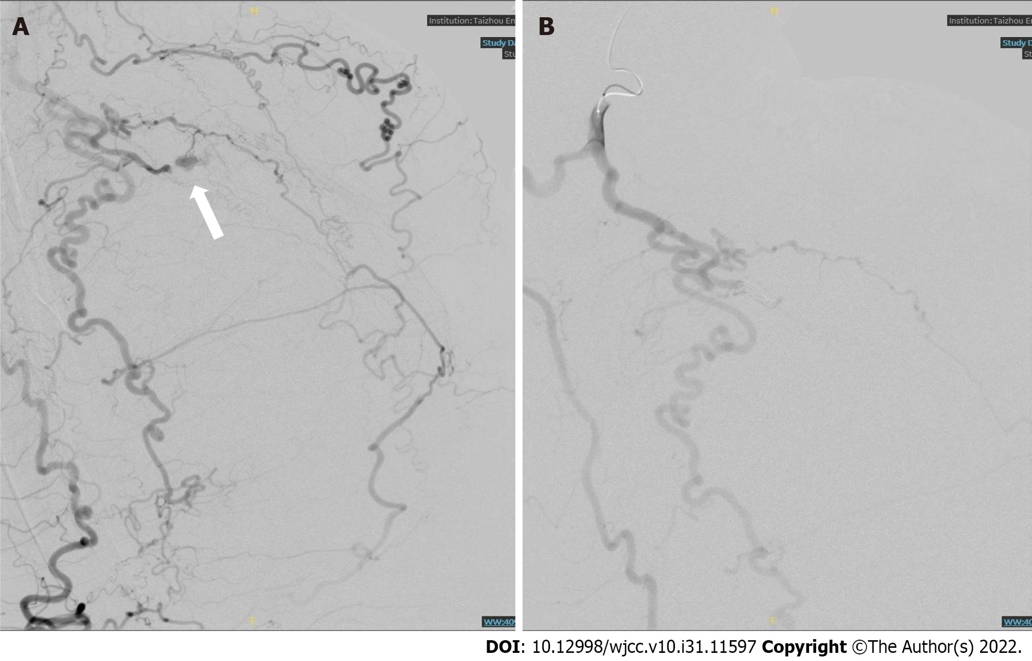
Figure 6 Right lower limb digital subtraction angiography and vascular embolization.
A: Digital subtraction angiography showed tortuous right popliteal and tibiofibular arteries and disordered tumor-like blood vessels. Contrast medium extravasation was seen in the local arterial branches in the right calf (white arrow); B: After vascular embolization, no contrast medium extravasation was found in the second angiography.
- Citation: Shen LP, Jin G, Zhu RT, Jiang HT. Hemorrhagic shock due to ruptured lower limb vascular malformation in a neurofibromatosis type 1 patient: A case report. World J Clin Cases 2022; 10(31): 11597-11606
- URL: https://www.wjgnet.com/2307-8960/full/v10/i31/11597.htm
- DOI: https://dx.doi.org/10.12998/wjcc.v10.i31.11597


