Copyright
©The Author(s) 2022.
World J Clin Cases. Nov 6, 2022; 10(31): 11597-11606
Published online Nov 6, 2022. doi: 10.12998/wjcc.v10.i31.11597
Published online Nov 6, 2022. doi: 10.12998/wjcc.v10.i31.11597
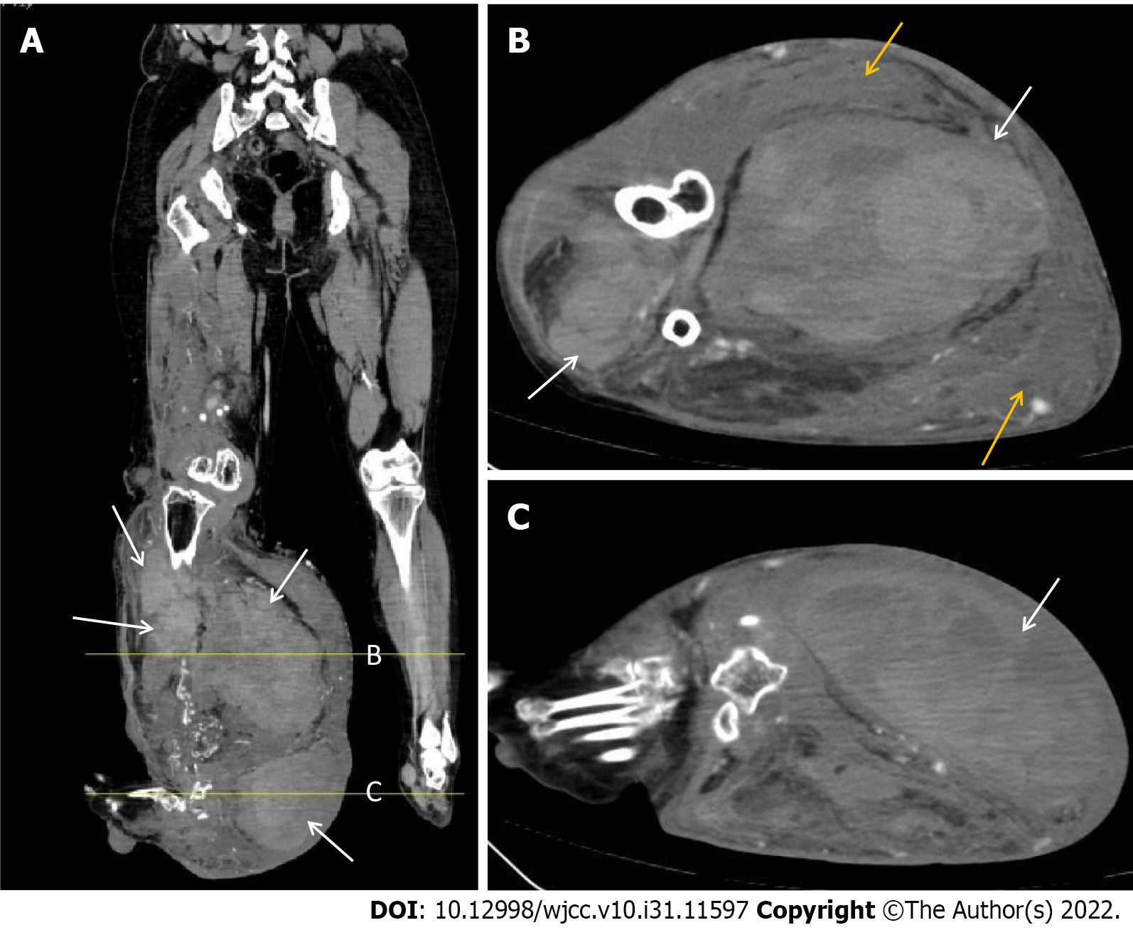
Figure 5 The soft tissue window of the right lower extremity computed tomography.
A: Sagittal view showed multiple large masses in the front and back of the right calf (white arrow, Lines A and B represent Figures A and B, respectively); B and C: Axial view showed multiple large masses in the front and back of the right calf and a subcutaneous hematoma behind the right calf (yellow arrow).
- Citation: Shen LP, Jin G, Zhu RT, Jiang HT. Hemorrhagic shock due to ruptured lower limb vascular malformation in a neurofibromatosis type 1 patient: A case report. World J Clin Cases 2022; 10(31): 11597-11606
- URL: https://www.wjgnet.com/2307-8960/full/v10/i31/11597.htm
- DOI: https://dx.doi.org/10.12998/wjcc.v10.i31.11597


