Copyright
©The Author(s) 2022.
World J Clin Cases. Nov 6, 2022; 10(31): 11549-11554
Published online Nov 6, 2022. doi: 10.12998/wjcc.v10.i31.11549
Published online Nov 6, 2022. doi: 10.12998/wjcc.v10.i31.11549
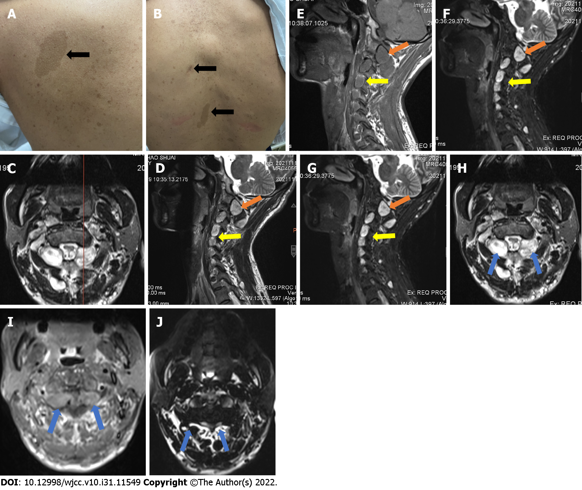
Figure 1 The physical signs and imaging data pre-operation.
A: Representative café-au-lait spots near the left scapula and on the back (B); C-J: Preoperative MRI images of the patient, showing conspicuous neoplasms in the upper cervical spine (yellow and blue arrows) and the presence of nodules at C2-7 (yellow arrows); C: At the position line of the sagittal and coronal planes, the neoplasm presents high signal on T1 (D, H), relatively low signal on T2 (E, I); signal enhancement showed high intensity in all nodules (F, J) images.
- Citation: Wang S, Ma JX, Zheng L, Sun ST, Xiang LB, Chen Y. Multiple bilateral and symmetric C1-2 ganglioneuromas: A case report. World J Clin Cases 2022; 10(31): 11549-11554
- URL: https://www.wjgnet.com/2307-8960/full/v10/i31/11549.htm
- DOI: https://dx.doi.org/10.12998/wjcc.v10.i31.11549


