Copyright
©The Author(s) 2022.
World J Clin Cases. Nov 6, 2022; 10(31): 11500-11507
Published online Nov 6, 2022. doi: 10.12998/wjcc.v10.i31.11500
Published online Nov 6, 2022. doi: 10.12998/wjcc.v10.i31.11500
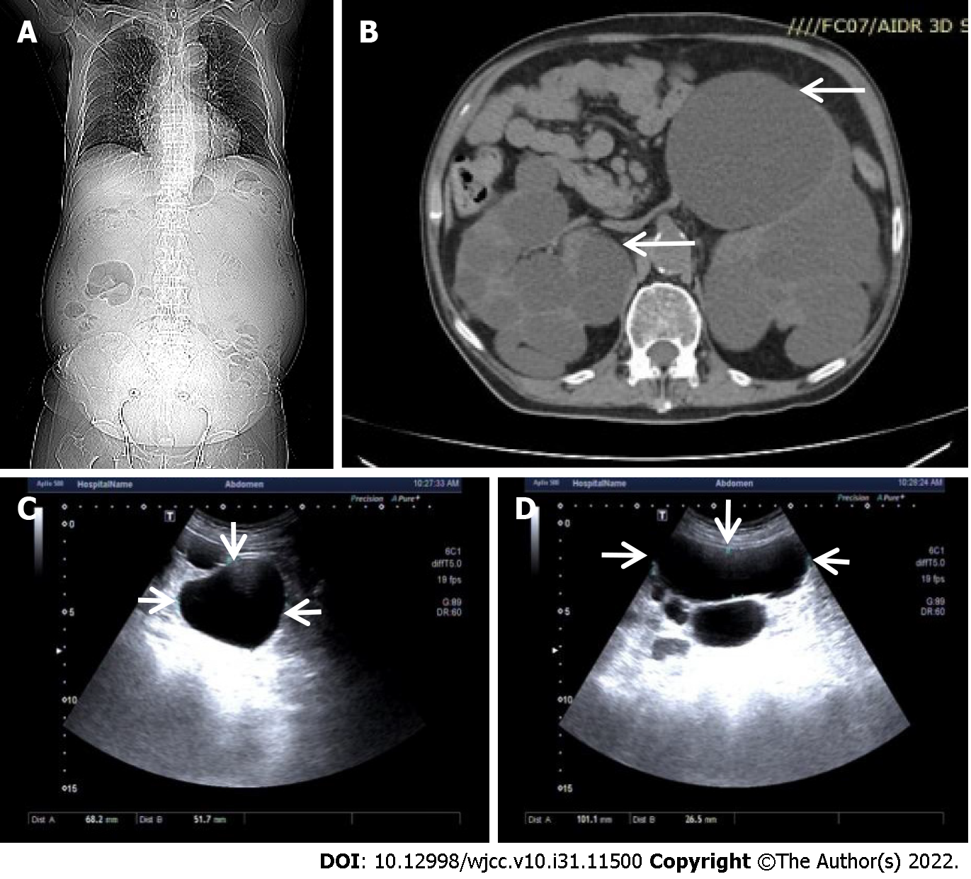
Figure 1 Representative images from computed tomography and renal ultrasonography.
A: Computed tomography imaging of the body; B: Representative image of multiple cysts on bilateral kidneys indicated by arrows; C: The largest cyst image on the right kidney indicated by arrows; D: The largest cyst image on the left kidney indicated by arrows.
- Citation: Zhou L, Tian Y, Ma L, Li WG. Tolvaptan ameliorated kidney function for one elderly autosomal dominant polycystic kidney disease patient: A case report. World J Clin Cases 2022; 10(31): 11500-11507
- URL: https://www.wjgnet.com/2307-8960/full/v10/i31/11500.htm
- DOI: https://dx.doi.org/10.12998/wjcc.v10.i31.11500


