Copyright
©The Author(s) 2022.
World J Clin Cases. Nov 6, 2022; 10(31): 11349-11357
Published online Nov 6, 2022. doi: 10.12998/wjcc.v10.i31.11349
Published online Nov 6, 2022. doi: 10.12998/wjcc.v10.i31.11349
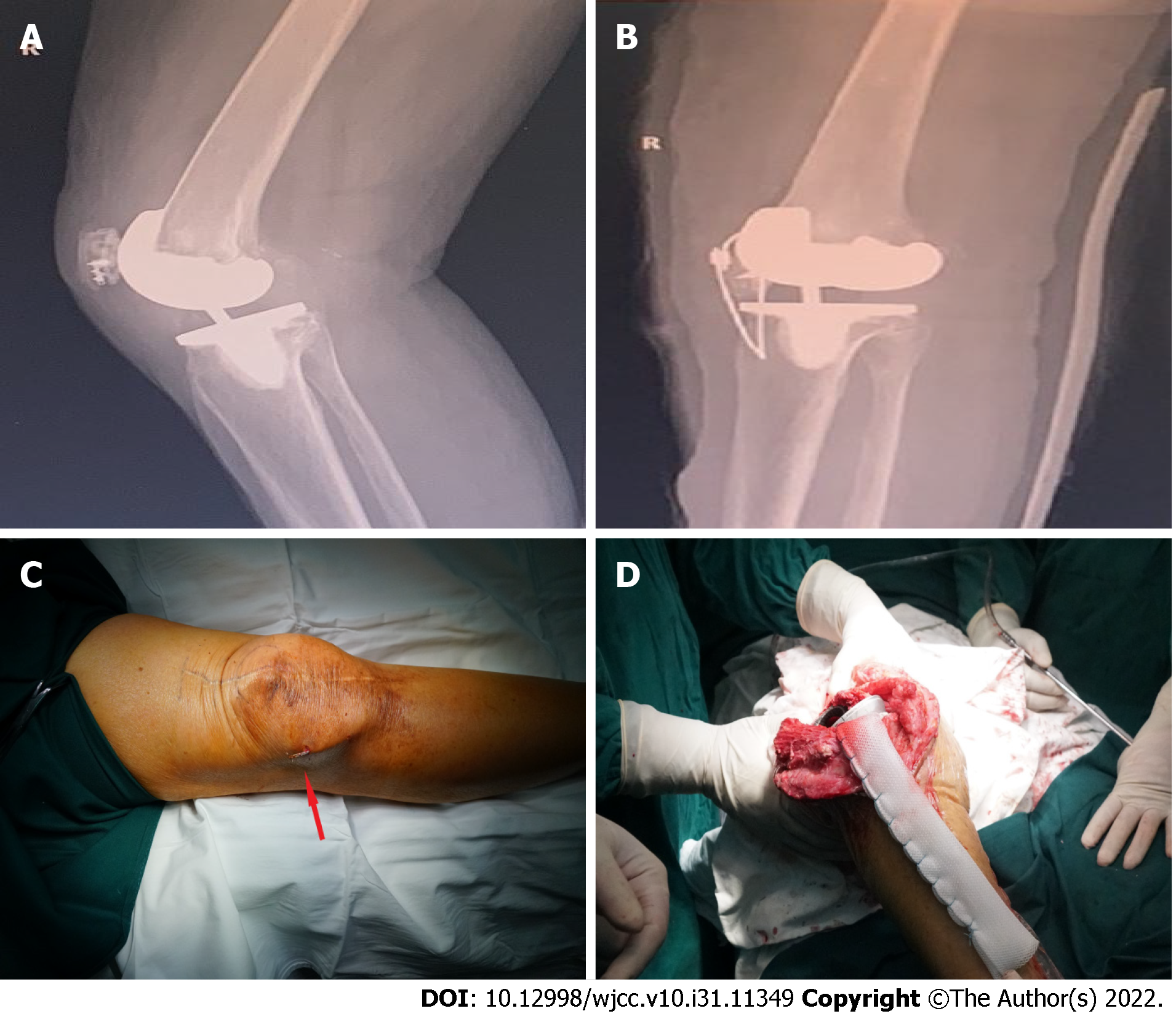
Figure 2 X-ray images.
A: X-ray image of the patient after suture anchoring treatment in another hospital; B: X-ray image after direct repair of patellar tendon fracture; C: The titanium cable in the knee joint was broken and the broken titanium cable punctured the skin to form a sinus; D: The patient was treated with Marlex mesh.
- Citation: Li TJ, Sun JY, Du YQ, Shen JM, Zhang BH, Zhou YG. Early patellar tendon rupture after total knee arthroplasty: A direct repair method. World J Clin Cases 2022; 10(31): 11349-11357
- URL: https://www.wjgnet.com/2307-8960/full/v10/i31/11349.htm
- DOI: https://dx.doi.org/10.12998/wjcc.v10.i31.11349


