Copyright
©The Author(s) 2022.
World J Clin Cases. Oct 26, 2022; 10(30): 11204-11209
Published online Oct 26, 2022. doi: 10.12998/wjcc.v10.i30.11204
Published online Oct 26, 2022. doi: 10.12998/wjcc.v10.i30.11204
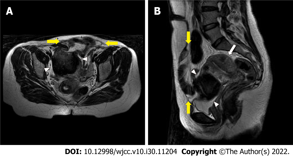
Figure 1 Pelvic magnetic resonance imaging shows thin skin and a fascial defect (yellow arrows) at the anterior pelvic wall.
The right rectus abdominis muscle is intact, but the left rectus abdominis muscle is atrophied. The subserosal uterine fibroid (white arrowheads) was located at the anterior of the uterus (white arrow). A: Axial T2-weighted image; B: Sagittal T2-weighted image.
- Citation: Park JW, Choi HY. Ventral hernia after high-intensity focused ultrasound ablation for uterine fibroids treatment: A case report. World J Clin Cases 2022; 10(30): 11204-11209
- URL: https://www.wjgnet.com/2307-8960/full/v10/i30/11204.htm
- DOI: https://dx.doi.org/10.12998/wjcc.v10.i30.11204


