Copyright
©The Author(s) 2022.
World J Clin Cases. Oct 26, 2022; 10(30): 11178-11184
Published online Oct 26, 2022. doi: 10.12998/wjcc.v10.i30.11178
Published online Oct 26, 2022. doi: 10.12998/wjcc.v10.i30.11178
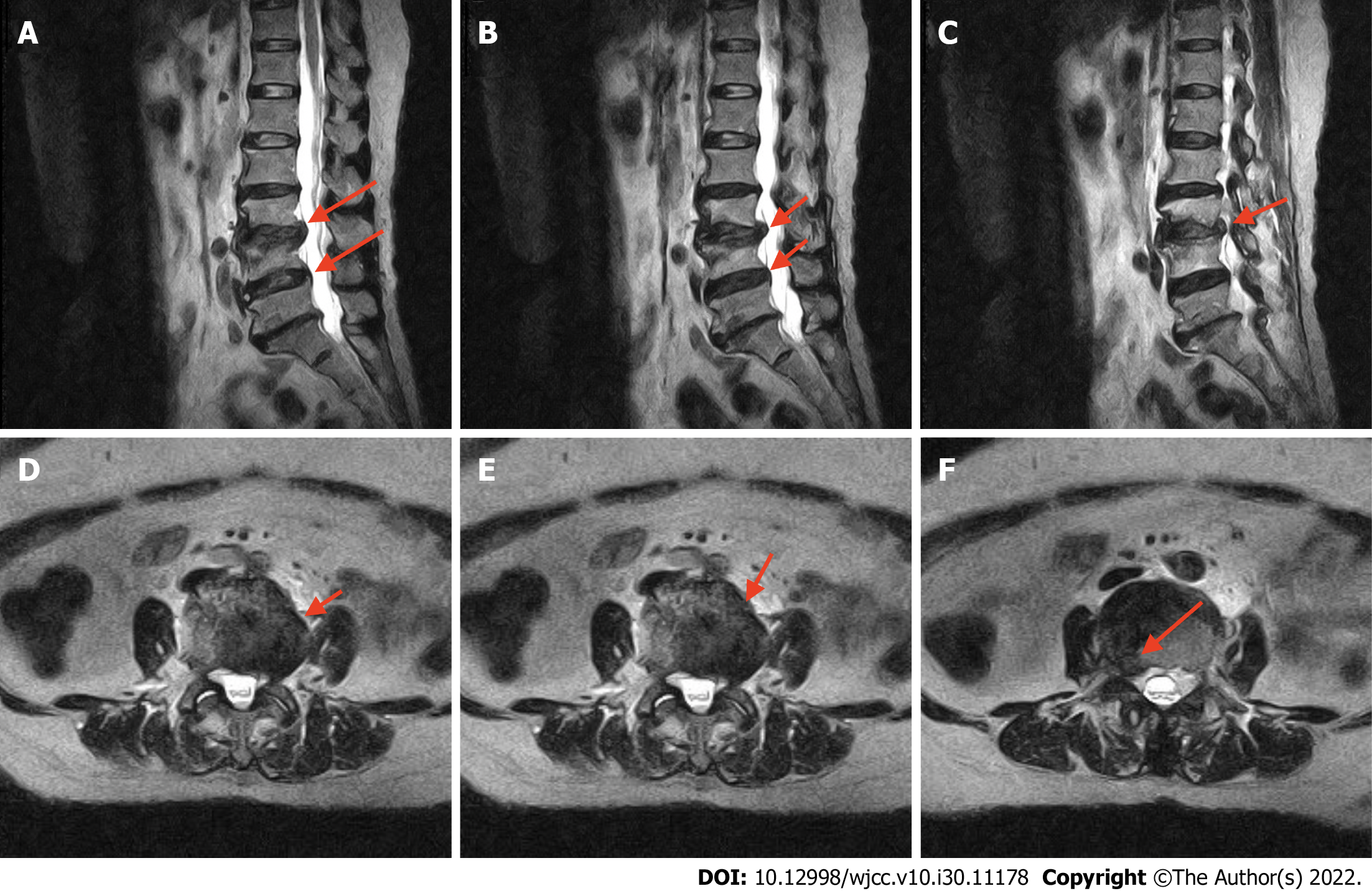
Figure 1 Preoperative T2-weighted magnetic resonance imaging.
The lateral and sagittal views of the collapsed disc height at the L2-3 and L3-4 Levels and a bulging disk compressing the thecal sac and right neural foramen are visible. A-C: Lateral views; D-F: Sagittal views.
- Citation: Yeh KL, Wu SH, Fuh CS, Huang YH, Chen CS, Wu SS. Cauda equina syndrome caused by the application of DuraSealTM in a microlaminectomy surgery: A case report. World J Clin Cases 2022; 10(30): 11178-11184
- URL: https://www.wjgnet.com/2307-8960/full/v10/i30/11178.htm
- DOI: https://dx.doi.org/10.12998/wjcc.v10.i30.11178


