Copyright
©The Author(s) 2022.
World J Clin Cases. Oct 26, 2022; 10(30): 11128-11138
Published online Oct 26, 2022. doi: 10.12998/wjcc.v10.i30.11128
Published online Oct 26, 2022. doi: 10.12998/wjcc.v10.i30.11128
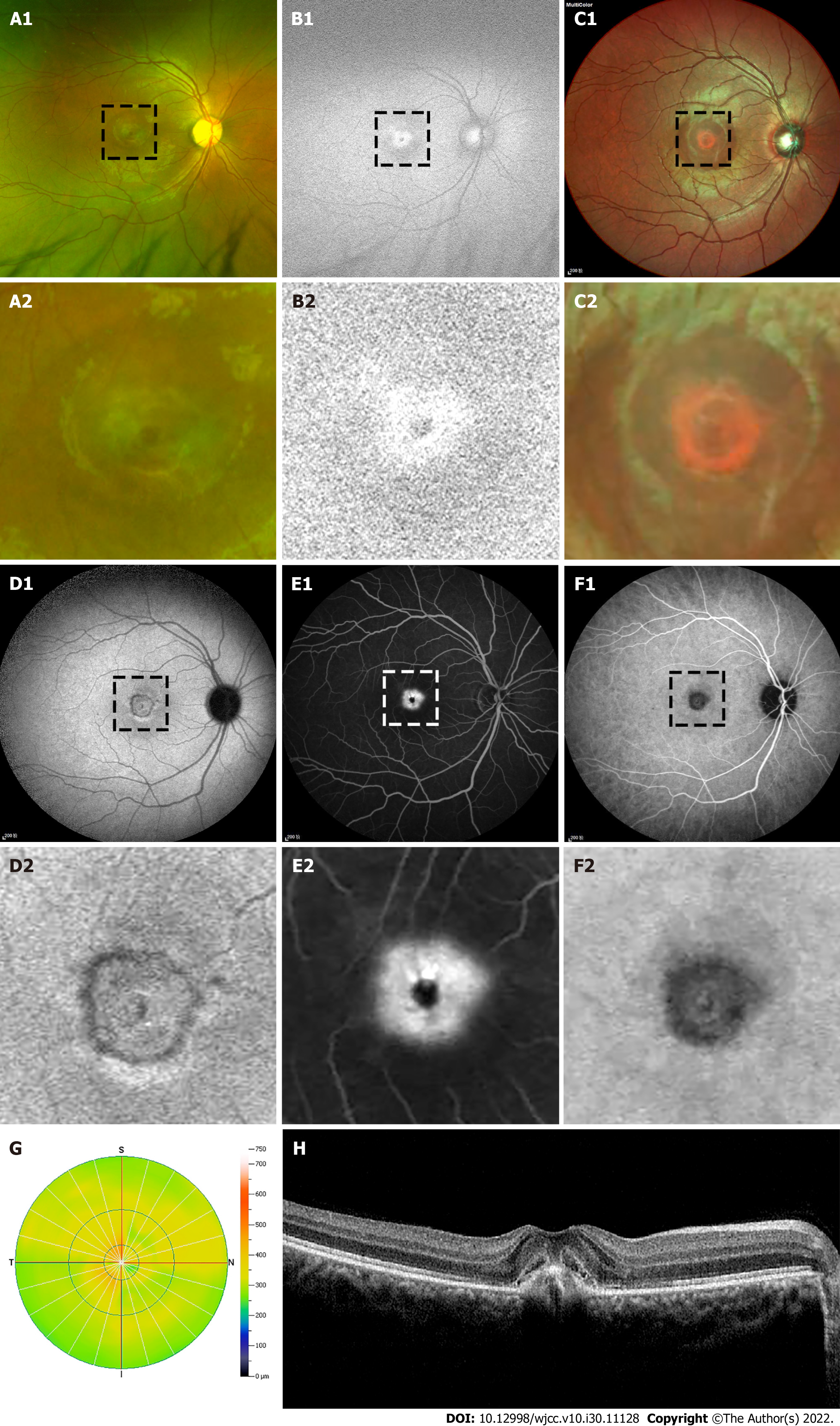
Figure 3 Retinal morphologic examinations.
A-D: Funduscopic and auto-fluorescence examination revealed a black shape punctuation abnormality surrounded with a ringlike margin lesion in the right eye; E and F: Angiography (FFA + ICGA) found a macular hyper-fluorescence leakage around a black shape punctation at right eye; G: Optical coherence tomography (OCT) revealed the macular fovea thickness increased; H: OCT showed the macular cystoid edema, retinal pigment epithelium layer breakdown and choroidal neovascularization in the right eye.
- Citation: Zhang X, Luo T, Mou YR, Jiang W, Wu Y, Liu H, Ren YM, Long P, Han F. Morphological and electrophysiological changes of retina after different light damage in three patients: Three case reports. World J Clin Cases 2022; 10(30): 11128-11138
- URL: https://www.wjgnet.com/2307-8960/full/v10/i30/11128.htm
- DOI: https://dx.doi.org/10.12998/wjcc.v10.i30.11128


