Copyright
©The Author(s) 2022.
World J Clin Cases. Oct 26, 2022; 10(30): 11101-11110
Published online Oct 26, 2022. doi: 10.12998/wjcc.v10.i30.11101
Published online Oct 26, 2022. doi: 10.12998/wjcc.v10.i30.11101
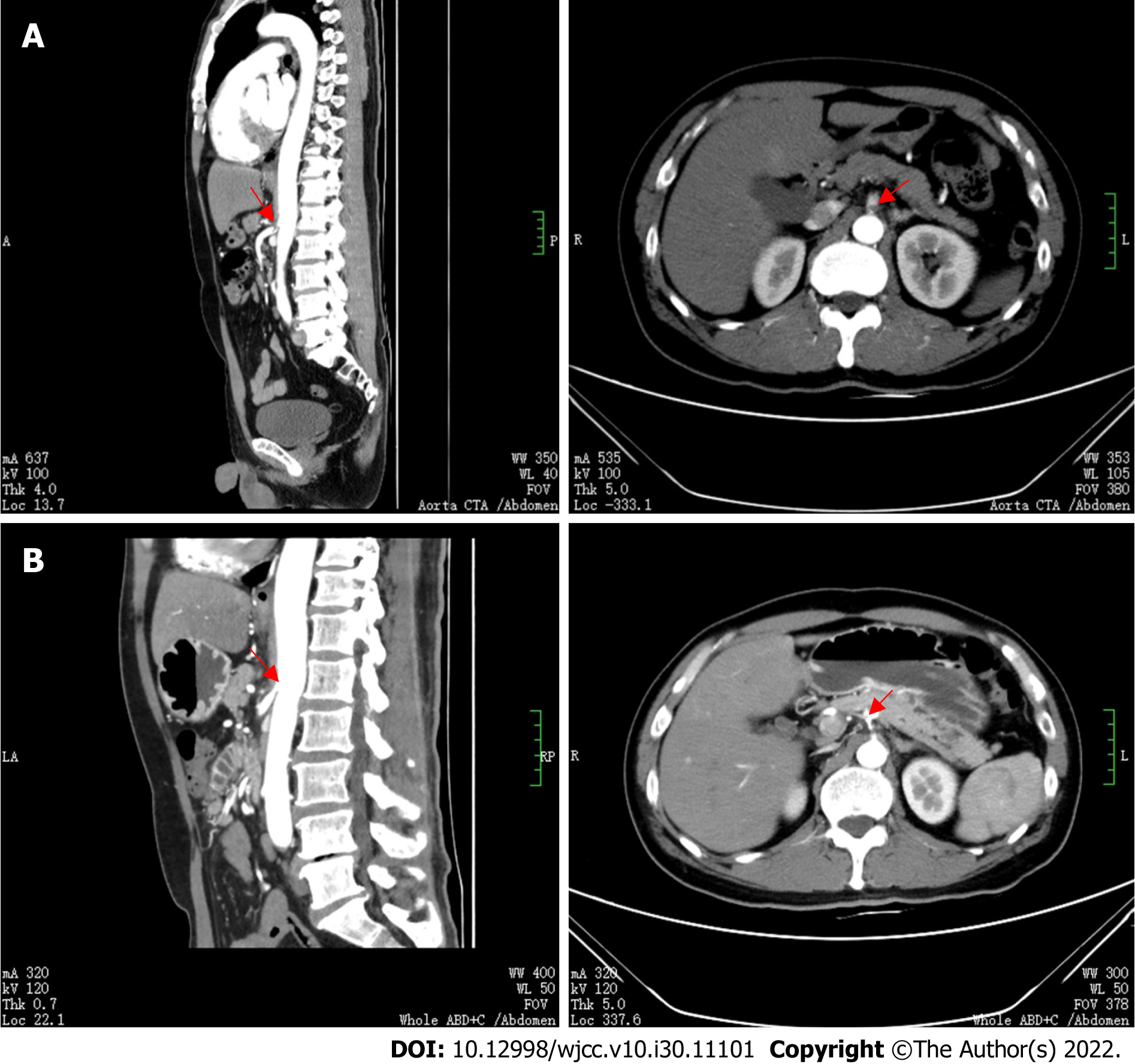
Figure 3 Abdominal aortic computed tomography angiography imaging examinations.
A: The proximal segment of the celiac trunk and superior mesenteric artery were embolized (as indicated by the red arrow), and the distal branch appeared small. The hepatic parenchyma around the gallbladder was enhanced in the arterial stage, with uneven local perfusion and a few calcified plaques in the abdominal aorta; B: Reperfusion of the proper hepatic artery, partial infarction of the spleen and cystic changes, blocked initial common pathway of the celiac trunk and superior mesenteric artery, but the embolization improved (as indicated by the red arrow), and obvious local stenosis.
- Citation: Bao XL, Tang N, Wang YZ. Severe Klebsiella pneumoniae pneumonia complicated by acute intra-abdominal multiple arterial thrombosis and bacterial embolism: A case report. World J Clin Cases 2022; 10(30): 11101-11110
- URL: https://www.wjgnet.com/2307-8960/full/v10/i30/11101.htm
- DOI: https://dx.doi.org/10.12998/wjcc.v10.i30.11101


