Copyright
©The Author(s) 2022.
World J Clin Cases. Oct 26, 2022; 10(30): 11101-11110
Published online Oct 26, 2022. doi: 10.12998/wjcc.v10.i30.11101
Published online Oct 26, 2022. doi: 10.12998/wjcc.v10.i30.11101
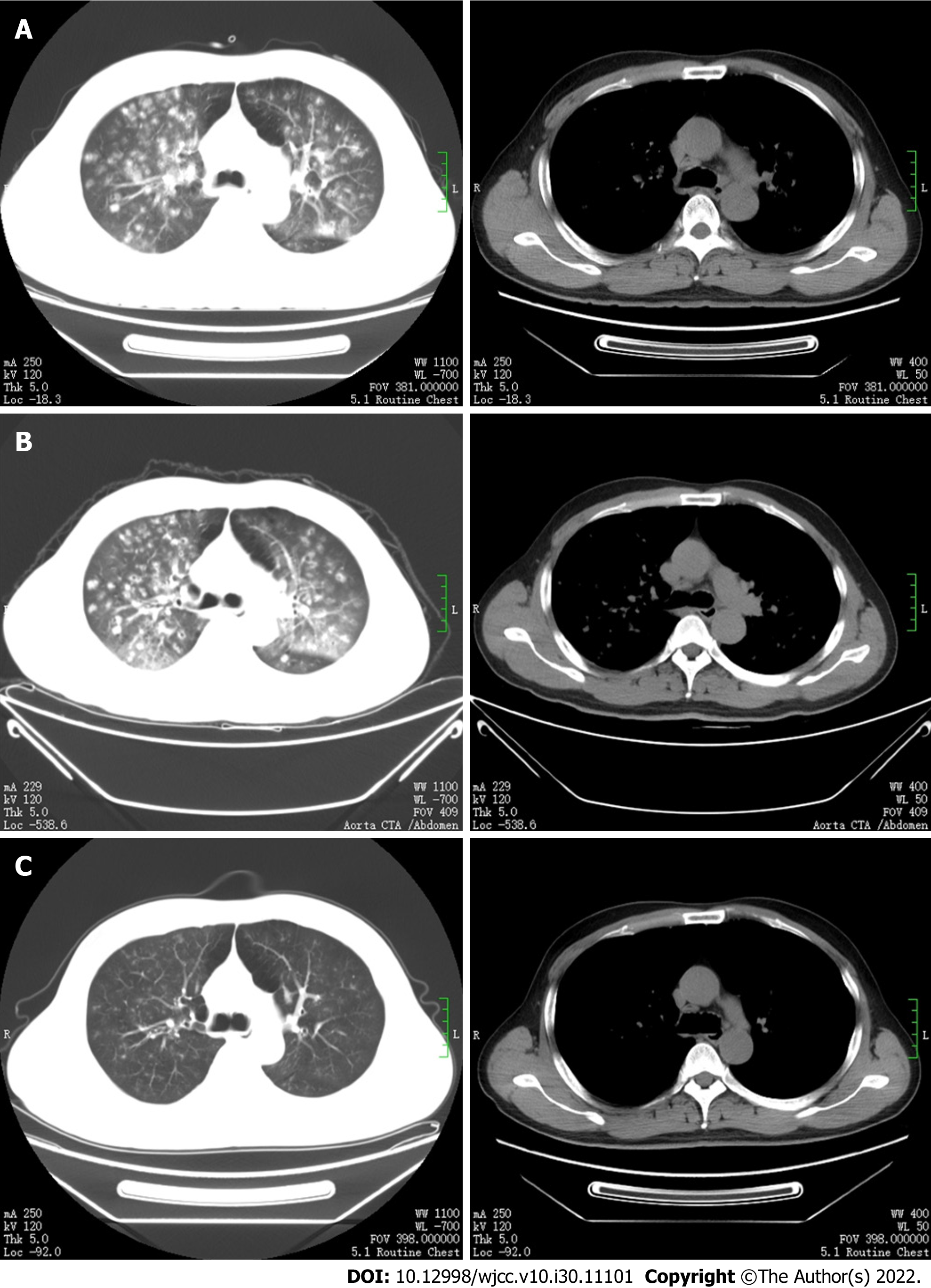
Figure 2 Chest computed tomography imaging examinations.
A: Multiple patchy, nodular, and flocculent high-density shadows in both lungs, with blurred edges and small voids in some lesions; B: The bilateral lung lesions increased, and the solid parts around some nodules increased, with reverse halo and trophovascular signs; C: The bilateral lung lesions were significantly absorbed and reduced compared with previous imaging.
- Citation: Bao XL, Tang N, Wang YZ. Severe Klebsiella pneumoniae pneumonia complicated by acute intra-abdominal multiple arterial thrombosis and bacterial embolism: A case report. World J Clin Cases 2022; 10(30): 11101-11110
- URL: https://www.wjgnet.com/2307-8960/full/v10/i30/11101.htm
- DOI: https://dx.doi.org/10.12998/wjcc.v10.i30.11101


